Main menu
Common skin conditions

NEWS
Join DermNet PRO
Read more
Quick links
Syringoma — extra information
Syringoma
Last Reviewed: December, 2024
Authors: Dr Amritpreet Singh, Dermatology Registrar ; A/Prof Amanda Oakley, Dermatologist, Waikato Hospital, NZ (2024)
Peer Reviewer: Dr Laura Wallace, Hutt Valley Hospital, NZ (2024)
Previous contributors: Hon A/Prof Amanda Oakley, Dermatologist (2003)
Reviewing dermatologist: Dr Ian Coulson
Edited by the DermNet content department
Introduction
Demographics and causes
Clinical features
Variation in skin types
Complications
Diagnosis
Differential diagnoses
Treatment
Outcome
What is a syringoma?
A syringoma is a benign adnexal tumour derived from the acrosyringium, which is the intraepidermal portion of an eccrine sweat duct. Syringomas are commonly found in clusters on and around the eyelids. They may also arise on the cheeks, scalp, axillae, umbilicus, chest, and genital regions.
Syringoma is derived from syrinx, the Greek word for tube or pipe. The Greek plural is syringomata. However, the use of ‘syringomas’ is more common.
In Friedman and Butler’s classification, four variants of syringomas are recognised:
- Localised syringomas
- Generalised multifocal or eruptive syringomas
- Syringomas associated with Down syndrome
- Familial syringomas.

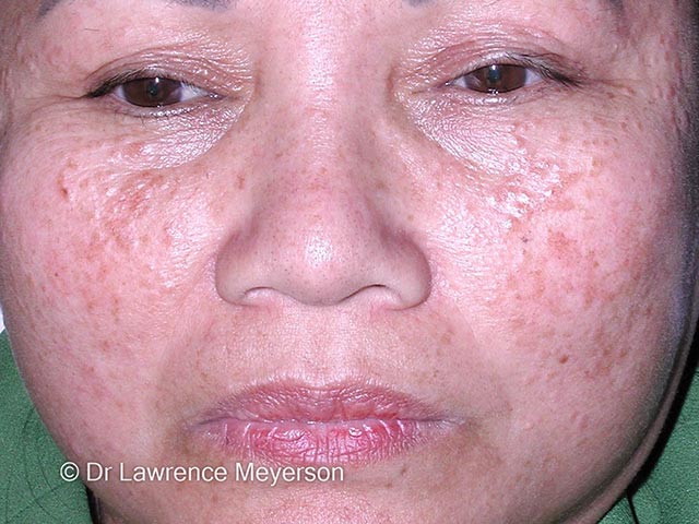
The typical 2-3mm flesh-coloured lid and cheek papules of syringomas (SY-patient1)

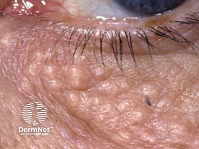
Close-up image of lid syringomas

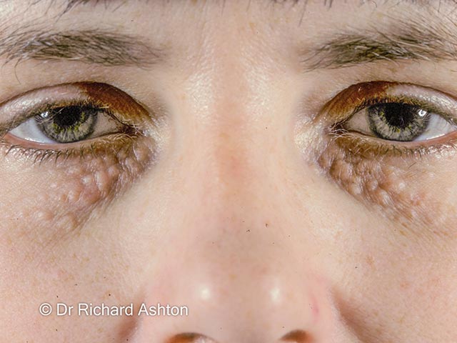
Symmetrical syringomas on the lids - they are larger than normal (SY-patient3)

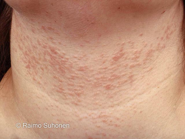
Lightly pigmented eruptive syringomas on the neck (SY-patient6)
Who gets and what causes syringomas?
Syringomas affect approximately 1% of the population. They are usually sporadic, appearing during or after adolescence. The male-to-female ratio is 1:2.
Familial syringomas are common, with an autosomal dominant pattern of inheritance. Inherited syringomas often arise before the onset of puberty and commonly affect the face.
Eyelid syringomas are associated with:
- Down syndrome: syringomas affect 18–39% of individuals with trisomy-21
- Marfan syndrome
- Ehlers–Danlos syndrome
- Brooke-Spiegler syndrome.
Eruptive syringomas are associated with:
- Asian ethnicity
- Darker skin types
- Down syndrome
- Nicolau–Balus syndrome*.
Vulvar syringomas have an incidence of 1:1100–1500, and their appearance may be precipitated by hormonal changes, such as during pregnancy.
Clear cell syringoma is a rare variant characterised by glycogen deposition and associated with diabetes mellitus.
*Nicolau–Balus syndrome (NBS) is a rare, autosomal dominant disorder that causes a group of deformities, including: generalised eruptive syringomata, atrophoderma vermiculata, and milia cysts.
What are the clinical features of syringomas?
Syringomas are firm, rounded dermal papules one to three millimetres in diameter.
- They are skin coloured, white, yellow or hyperpigmented.
- They may appear translucent or cystic.
- They may be solitary or occur in clusters, often symmetrically distributed.
- The lower eyelids and cheeks are the most common sites.
Eruptive syringomas appear as crops of hyperpigmented papules widespread across the trunk, chest, abdomen, upper extremities, female genitalia (where they are found on the labia majora) or rarely occur on the penile shaft.
Syringomas are usually asymptomatic but may cause itching associated with sweating or when located on the vulva.
How do clinical features vary in differing types of skin?
In people with skin of colour, syringomas are yellowish, hypopigmented, or hyperpigmented papules.
What are the complications of syringoma?
Syringomas are benign. Their significance is primarily cosmetic.
Syringomas associated with Down syndrome may calcify (calcinosis cutis).
How is syringoma diagnosed?
A syringoma is often diagnosed clinically due to its typical appearance.
A skin biopsy of syringoma reveals a normal epidermis, multiple small ducts, and epithelial cords in the dermis. Two rows of flattened epithelial cells line the ducts, the outer layer bulging outward to create a comma-like tail resembling a tadpole. The surrounding stroma is sclerotic.
Clear cell syringomas have a clear cytoplasm of the epithelial cells due to their increased levels of glycogen. When confirmed via biopsy, further evaluation for glucose intolerance is recommended due to their association with diabetes mellitus.
A full skin thickness biopsy is necessary to exclude microcystic adnexal carcinoma, which has similar features but infiltrates deep dermis and subcutaneous tissue.
What is the differential diagnosis for syringoma?
The differential diagnosis for syringoma includes:
- Xanthelasma
- Trichoepithelioma
- Milium
- Fox-Fordyce disease
- Microcystic adnexal carcinoma
- Basal cell carcinoma.
What is the treatment for syringoma?
Syringomas are benign and do not require intervention. Treatment for cosmetic reasons aims to reduce tumour visibility with minimal scarring, but the results are often suboptimal.
Treatment options include:
- Topical atropine sulphate to reduce pruritus, if present
- Long-term tretinoin cream to improve the appearance
- Laser treatment with CO2 or erbium YAG laser
- Electrosurgery
- Surgical excision.
What is the outcome for syringomas?
Spontaneous regression of syringoma is rare.
Syringomas can recur after superficial treatment as they penetrate the deep dermis. Hypertrophic or atrophic scarring and post-inflammatory pigment changes are uncommon complications of treatment, particularly in people with darker skin types.
Bibliography
- Soler-Carrillo J, Estrach T, Mascaró JM. Eruptive syringoma: 27 new cases and review of the literature. J Eur Acad Dermatol Venereol. 2001;15(3):242-246. doi:10.1046/j.1468-3083.2001.00235.x. PubMed
- Friedman SJ, Butler DF. Syringoma presenting as milia. J Am Acad Dermatol. 1987 Feb;16(2 Pt 1):310-4. doi: 10.1016/s0190-9622(87)70041-3. PMID: 3819065. PubMed
- Ghanadan A, Khosravi M. Cutaneous syringoma: a clinicopathologic study of 34 new cases and review of the literature. Indian J Dermatol. 2013;58(4):326. doi:10.4103/0019-5154.113956. PubMed Central
- Asha GS, Aithal SS, Revathi TN, Shilpa K. Vulvar syringoma - An uncommon presentation. Indian J Sex Transm Dis AIDS. 2022;43(1):93-94. doi:10.4103/ijstd.ijstd_135_20. PubMed Central
- Miranda JJ, Shahabi S, Salih S, Bahtiyar OM. Vulvar syringoma, report of a case and review of the literature. Yale J Biol Med. 2002;75(4):207-210. PubMed
