Main menu
Common skin conditions

NEWS
Join DermNet PRO
Read more
Quick links
Steatocystoma multiplex — extra information
Steatocystoma multiplex
Author: Dr Amanda Oakley Dermatologist, Hamilton New Zealand. Updated February 2014. DermNet Revision September 2021
Introduction Demographics Causes Clinical features Complications Diagnosis Differential diagnoses Treatment
What is steatocystoma multiplex?
Steatocystoma multiplex is a rare inherited disorder in which numerous hamartomatous malformations of the pilosebaceous duct junction (hair follicle unit) develop at puberty.
If a single cyst of this type is found, it is called steatocystoma simplex.
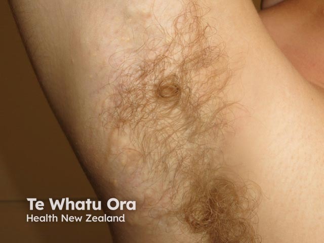
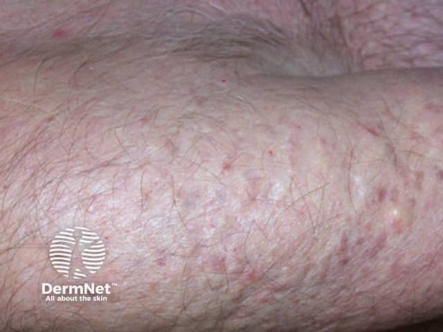
Steatocystoma multiplex
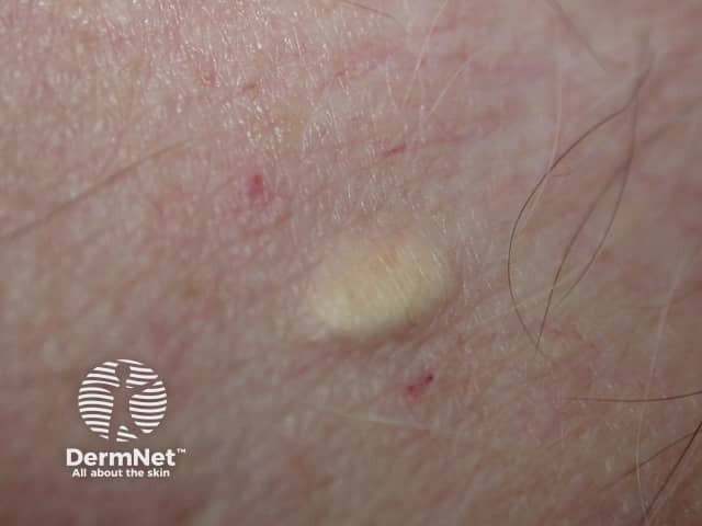
Who gets steatocystoma multiplex?
In steatocystoma multiplex, the tendency to develop cysts is inherited in an autosomal dominant fashion, so one parent can be expected to also have steatocystoma multiplex. It may also occur sporadically. Both males and females may be affected.
What is the cause of steatocystoma multiplex?
Familial steatocystoma multiplex is associated with mutations in the keratin 17 gene (K17). This gene is responsible for a keratin protein found in the nail bed, hair follicles and sebaceous glands. Mutations in K17 may also result in one form of pachyonychia congenita, in which cysts, plantar keratoderma and natal teeth are common.
Steatocystomas are thought to come from an abnormal lining of the passageway to the oil glands (sebaceous duct).
The onset at puberty is presumably due to hormonal stimulus of the pilosebaceous unit.
What are the clinical features of steatocystoma multiplex?
The cysts of steatocystoma multiplex most often arise on the chest and may also occur on the abdomen, upper arms, armpits and face. In some cases cysts may develop all over the body.
The cysts are mostly small (2-20 mm) but they may be several centimetres in diameter. They tend to be soft to firm semi-translucent bumps, and contain an oily, yellow liquid. Sometimes a small central punctum can be identified and they may contain one or more hairs (eruptive vellus hair cysts).
Localised, generalised, facial, acral, and suppurative types of steatocystoma multiplex have been described. Isolated steatocystoma of the vulva and scrotum can develop late in life as a sporadic condition not inherited (see Vulval cysts).
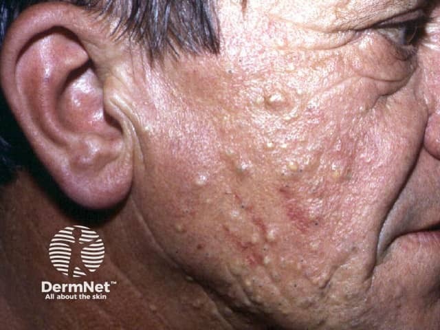
Steatocystoma multiplex
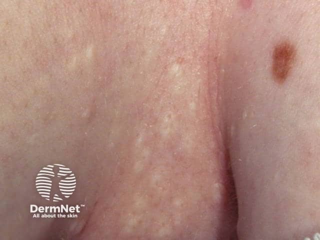
Steatocystoma multiplex
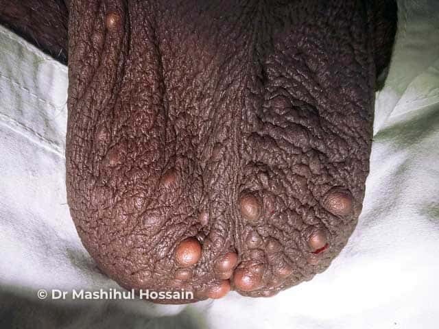
Steatocystoma multiplex
What are the complications of steatocystoma multiplex?
- Psychosocial impact on quality of life
- Inflammation and scarring
How is steatocystoma multiplex diagnosed?
Skin biopsy of a cyst will typically show a thin-walled dermal cyst with a sebaceous gland in the cyst wall and lanugo hair within the cyst lumen.
What is the differential diagnosis of steatocystoma multiplex?
What is the treatment for steatocystoma multiplex?
- Individual cysts can be removed surgically. In most cases, small incisions (cuts into the skin) allow the cyst and its contents to be extracted through the opening. If it is tethered to the underlying skin, excision biopsy may be necessary.
- Cysts can also be removed by laser, electrosurgery or cryotherapy.
- Inflammation can be reduced with oral antibiotics
- Oral isotretinoin is not curative but may temporarily shrink the cysts and reduce inflammation.
References
- Georgakopoulos JR, Ighani A, Yeung J. Numerous asymptomatic dermal cysts: diagnosis and treatment of steatocystoma multiplex. Can Fam Physician. 2018;64(12):892–9. Journal
- Kamra HT, Gadgil PA, Ovhal AG, Narkhede RR. Steatocystoma multiplex-a rare genetic disorder: a case report and review of the literature. J Clin Diagn Res. 2013;7(1):166–8. doi:10.7860/JCDR/2012/4691.2698 PubMed Central
On DermNet
Other websites
- Steatocystoma multiplex — Medscape Reference
- Steatocystoma multiplex #184500 — OMIM
