Main menu
Common skin conditions

NEWS
Join DermNet PRO
Read more
Quick links
Sebaceous carcinoma — extra information
Sebaceous carcinoma
Author: Vanessa Ngan, Staff Writer, 2013. Updated by A/Prof Amanda Oakley, Dermatologist, Hamilton, New Zealand, January 2018. DermNet Update July 2021
Introduction Demographics Causes Diagnosis Differential diagnoses Treatment Outcome
What is sebaceous carcinoma?
Sebaceous carcinoma is a rare aggressive skin cancer arising from a sebaceous gland. Sebaceous carcinoma is sometimes called sebaceous gland adenocarcinoma.
Who gets sebaceous carcinoma?
Periocular sebaceous carcinoma affects adults. It occurs more frequently in Asian populations than Caucasians and is more common in women than in men, particularly those around 60 to 80 years of age.
Extraocular sebaceous carcinomas occur mainly in older adults and without predilection for male or female.
Patients with Muir-Torre syndrome (a predisposing genetic syndrome) may be diagnosed at a younger age.
What causes sebaceous carcinoma?
Sebaceous glands are small glands connected to hair follicles in the skin. They are located in any hair-bearing region of the body but are most numerous on the skin of the scalp and face. The glands are responsible for producing sebum which is an oily substance that keeps hair and skin moisturised.
The exact cause of sebaceous carcinoma is unclear. The following have been reported to possibly increase the risk of these tumours:
- Underlying Muir-Torre or Lynch syndrome
- Previous radiation therapy to the area for a variety of benign and malignant conditions, such as retinoblastoma
- History of oral thiazide diuretic use
- Mutations to the tumour suppressor gene p53
- Immunosuppression.
What are the clinical features of sebaceous carcinoma?
Periocular sebaceous carcinoma
Sebaceous carcinoma most commonly develops from the meibomian glands of the eye which are located mostly in the upper but also in the lower eyelids.
- Small, erythematous or yellowish, firm, deep-seated, slowly enlarging nodule on the upper eyelid.
- Lesions occur on the upper eyelid 2–3 times more commonly than on the lower eyelid.
- The lesion is often mistaken for chalazion (a benign inflammation of the meibomian gland).
- As the carcinoma grows it may spread onto the conjunctiva, where it can be mistaken for keratoconjunctivitis or blepharoconjunctivitis.
- In advanced cases, the spread of the lesion may lead to both upper and lower lid lesions and cause loss of eyelashes (madarosis), ulceration, and distorted vision.
- Undiagnosed or late diagnoses can result in metastases (spread) to lymph nodes and parotid glands.
Extraocular sebaceous carcinoma
Extraocular sebaceous carcinoma accounts for about 25% of sebaceous carcinomas. These tumours mostly occur around the head and the neck. Other sites where these tumours have been found include the genitals, ear canal, breasts, trunk, and oral cavity.
The clinical presentation of extraocular lesions is as a non-specific tumour; they typically appear as a pink to a yellow-red nodule of varying sizes.
Extraocular sebaceous carcinoma
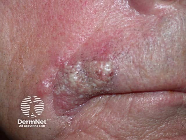
Sebaceous carcinoma
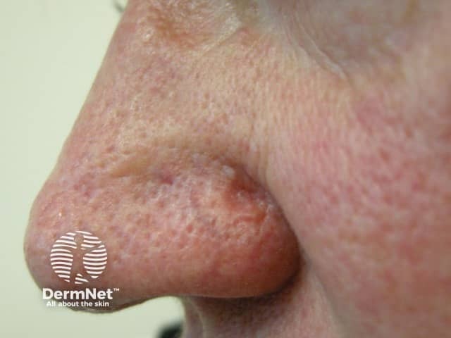
Sebaceous carcinoma
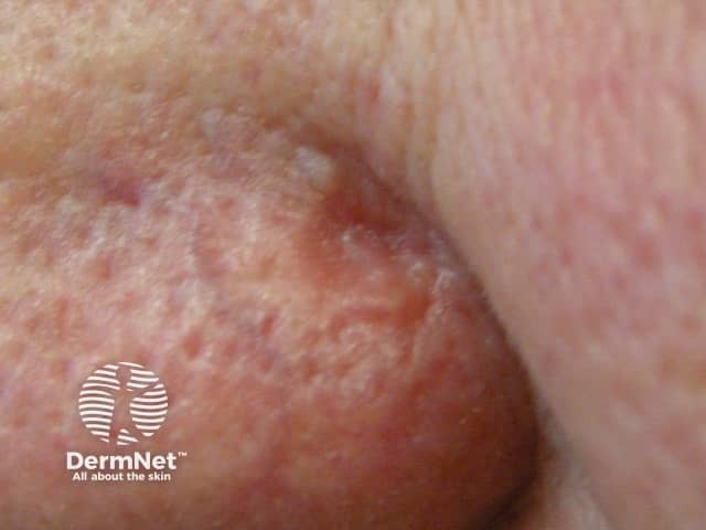
Sebaceous carcinoma
How is sebaceous carcinoma diagnosed?
Sebaceous carcinoma may be suspected clinically. Dermoscopy may reveal typical irregular yellowish lobules and irregular linear blood vessels.
Definitive diagnosis is based upon patient history, adequate surgical biopsy and the combined knowledge of a pathologist, ophthalmologist and dermatologist. [see Sebaceous carcinoma pathology]
Immunohistochemistry stains are routinely used to determine the likelihood of Muir-Torre syndrome.
Sebaceous carcinoma dermoscopy
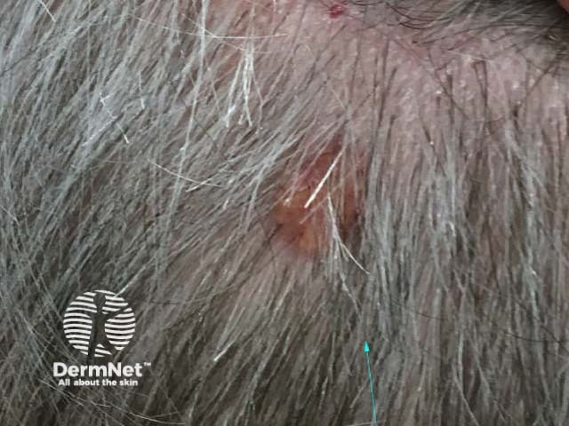
Clinical view
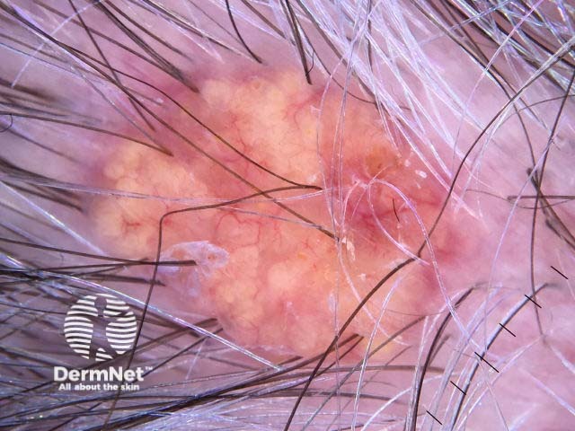
Dermoscopy view
What is the differential diagnosis of sebaceous carcinoma?
Conditions/diseases that mimic periocular sebaceous carcinoma
- Chalazion
- Hordeolum (stye)
- Pyogenic granuloma / reactive haemangioma
- Granulomatous inflammation from sarcoidosis, syphilis or tuberculosis
Other eyelid tumours
What are the complications of sebaceous carcinoma?
- Delayed diagnosis - diagnosis of periocular sebaceous carcinoma is often delayed by months to years (mean 1–3 years)
- Metastatic disease
- Recurrence - approximately 30% of sebaceous carcinomas recur after resection
How is sebaceous carcinoma treated?
Radical surgical excision with frozen section control by either a standard method or Mohs micrographic surgery is the most common and effective method of treatment.
Radiation therapy should only be used in patients unable or unwilling to undergo surgery.
What is the outcome for sebaceous carcinoma?
Sebaceous carcinoma is an aggressive and potentially dangerous tumour that can lead to significant morbidity and mortality. The overall mortality rate is 5–10% because of inherent tumour factors, or delayed diagnosis and treatment.
Factors for a poorer prognosis include delay in diagnosis of greater than 6 months, tumour diameter greater than 1 cm, and both upper and lower eyelid involvement.
Bibliography
- Eiger-Moscovich M, Eagle RC Jr, Shields CL, et al. Muir-Torre syndrome associated periocular sebaceous neoplasms: screening patterns in the literature and in clinical practice. Ocul Oncol Pathol. 2020;6(4):226–37. doi:10.1159/000504984. Journal
- Kibbi N, Worley B, Owen JL, et al. Sebaceous carcinoma: controversies and their evidence for clinical practice. Arch Dermatol Res. 2020;312(1):25–31. doi:10.1007/s00403-019-01971-4. PubMed
On DermNet
- Muir-Torre syndrome
- Blepharitis
- Skin surgery
- Skin cancer
- Skin lesions, tumours and cancers
- Hair follicle tumours
Other websites
- Sebaceous carcinoma — American Academy of Dermatology
- Dermatological Manifestations of Sebaceous Carcinoma — Medscape Reference
- Sebaceous carcinoma — Medscape Reference
