Main menu
Common skin conditions

NEWS
Join DermNet PRO
Read more
Quick links
Pemphigus foliaceus pathology — extra information
Autoimmune/autoinflammatory Diagnosis and testing
Pemphigus foliaceus pathology
Author: Dr Ben Tallon, Dermatologist/Dermatopathologist, Tauranga, New Zealand, 2010.
Histology Special stains Histological variants Differential diagnoses
Pemphigus foliaceus is grouped within the intraepidermal blistering disorders.
Histology of pemphigus foliaceus
The low power view of the histology of pemphigus foliaceus is of a superficial epidermal blistering process. The low power clues include loss of the stratum corneum, increased prominence of the granular layer, or visible superficial epidermal separation with blister formation (Figure 1). At higher magnification subtle acanthloysis and spongiosis can be seen within the stratum granulosum, extending into the stratum corneum (Figures 2 and 3). This can form separation within the superficial epidermis, or as mentioned above, lead to complete loss of the stratum corneum. The prominent granular layer is seen as hyperchromasia of the nuclei within dyskeratotic cells in this layer, similar to the grains seen in Dariers disease (Figures 4 and 5).
In the dermis there is a predominantly superficial lymphocytic infiltrate with scattered eosinophils (Figure 3). Neutrophils may be more common in the IgA subtype.
If there is clinical suspicion for pemphigus folicaceous but little to see on first inspection, remember to assess the hair follicles, as early changes may be seen here.
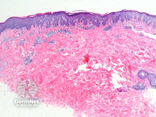
Figure 1
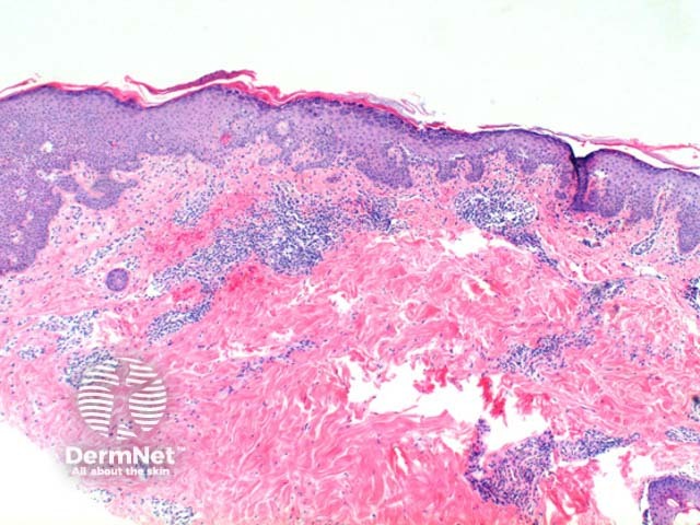
Figure 2
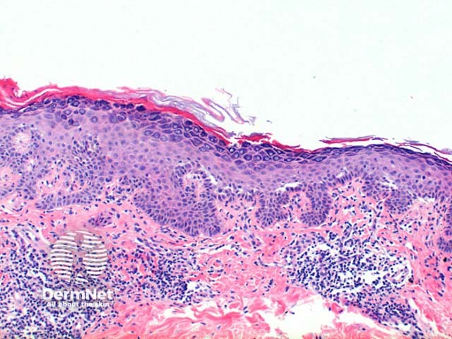
Figure 3
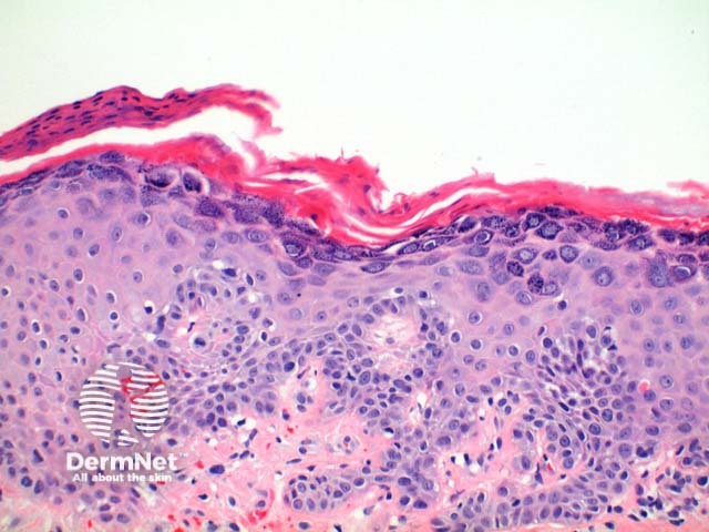
Figure 4
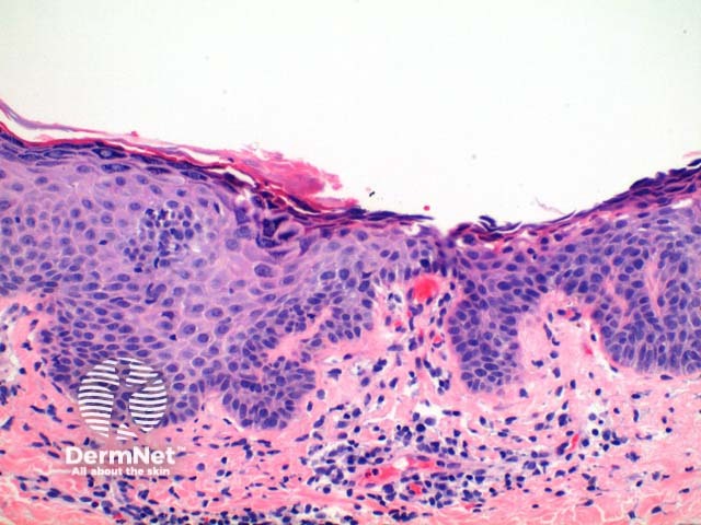
Figure 5
Special stains in pemphigus foliaceus
Direct immunofluorescence is a critical component of the workup, but requires a separate specimen transported in appropriate media. A positive result is the presence of intercellular IgG and C3 predominantly within the superficial layers of the epidermis (Figure 6).
Pemphigus foliaceus – direct immunofluorescence
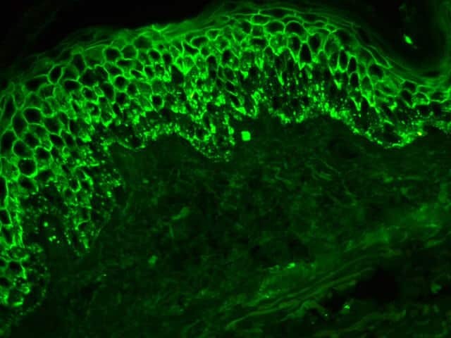
Figure 6
Histological variants of pemphigus foliaceus
Pemphigus erythematosus: In this variant the histology is that of pemphigus foliaceus, with the additional immunoflouorescence finding of basal membrane IgG +/- C3 in addition to the intercellular distribution. Antinuclear antibody studies are also frequently positive.
IgA pemphigus: Typically there is a more prominent subcorneal neutrophilic infiltrate forming pustules. When there is only minor vesicle formation the clue to this diagnosis is in the predominance of neutrophils. Immunofluorescence show intercellular IgA deposition.
Differential diagnosis of pemphigus foliaceus
Pemphigus vulgaris: In this related condition intraepidermal acantholysis is seen within the lower suprabasal epidermis, with matching direct immunofluorescence at this level.
Acantholytic actinic keratosis: In this condition there is prominent atypia of the keratinocytes, with acantholysis frequently present through all layers of the involved epidermis. Discrimination may be difficult when pemphigus foliaceus is seen on markedly solar damaged skin.
Staphylococcal scalded skin syndrome: In this acute condition there is widespread subcorneal separation, typically with complete absence of the stratum corneum without prominence of the granular layer and minimal inflammatory infiltrate.
Herpetiform pemphigus: This should be considered when the clinical history is suggestive of dermatitis herpetiforms. The histology may show subcorneal separation and superficial epidermal intercellular IgG deposition simulating pemphigus foliaceus. Typically cases demonstrate more prominent eosinophilic spongiosis with intraepidermal blister formation and occasionally subcorneal neutrophilic pustulation.
References
- Skin Pathology (2nd edition, 2002). Weedon D
- Pathology of the Skin (3rd edition, 2005). McKee PH, J. Calonje JE, Granter SR
On DermNet
