Main menu
Common skin conditions

NEWS
Join DermNet PRO
Read more
Quick links
Idiopathic facial aseptic granuloma — extra information
Idiopathic facial aseptic granuloma
Last reviewed: April 2023
Authors: Dr Anne-Marie Aubin, Resident; Dr Kirsty Wark, Dermatology advanced trainee; Dr John Relic, Consultant dermatologist, John Hunter Hospital, Australia (2023)
Reviewing dermatologist: Dr Ian Coulson
Edited by the DermNet content department
Introduction Demographics Causes Clinical features Variation in skin types Complications Diagnosis Differential diagnoses Treatment Prevention Outcome
What is idiopathic facial aseptic granuloma?
Idiopathic facial aseptic granuloma (IFAG) is a rare, painless, self-limiting, solitary nodule which affects children.
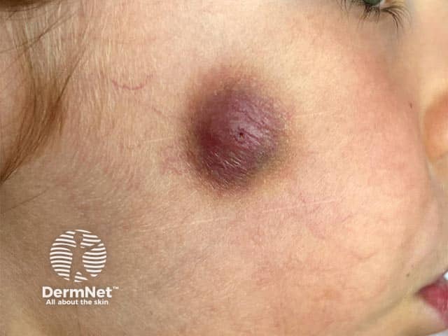
It is reminiscent of an acne nodule and usually heals without scarring. It typically presents on the cheek or eyelids as a red-violaceous papule or nodule. Multiple lesions are uncommon.

Violaceous plaque on the cheek due to idiopathic facial aseptic granuloma

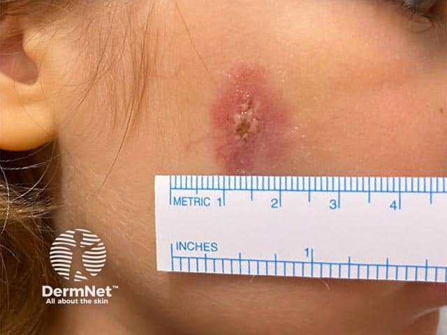
Violaceous indurated plaque with central ulceration in idiopathic facial aseptic granuloma

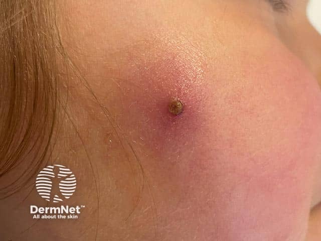
Violaceous plaque with some overlying crusting due to idiopathic facial aseptic granuloma

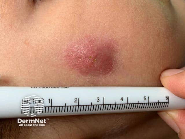
Indurated plaque over the cheek in a child with idiopathic facial aseptic granuloma
Who gets idiopathic facial aseptic granuloma?
Idiopathic facial aseptic granuloma (IFAG) occurs exclusively in young children and adolescents, with a mean age of 4 years at presentation.
It appears to be twice as common in females than in males.
What causes idiopathic facial aseptic granuloma?
The pathogenesis of IFAG remains unclear but is thought to be a type of childhood granulomatous rosacea. This theory originates from its association with chalazions, telangiectasias, and localisation to the eyelids, fitting the rosacea spectrum.
Other theories include a reactive process to insect bites or trauma, or an embryonic remnant.
No clear risk factors or associated clinical features have been described.
What are the clinical features of idiopathic facial aseptic granuloma?
- Painless red-violaceous solitary papule or nodule.
- Most lesions arise in a region formed by the corner of the mouth, the earlobe, and the outer angle of the eye.
- May be soft or firm; often described to have an ‘elastic’ consistency.
- Variable size (3–30 mm).
- Local incision of a nodule can result in the discharge of pus or blood (with negative cultures unless there is superinfection).
How do clinical features vary in differing types of skin?
Further research is needed to describe features of idiopathic facial aseptic granuloma in darker Fitzpatrick skin phototypes and races.
What are the complications of idiopathic facial aseptic granuloma?
Complications of idiopathic facial aseptic granuloma (IFAG) are rare.
However, IFAG increases the risk of developing rosacea in the future, particularly ocular rosacea. Thus, complications of ocular rosacea may occur, such as blepharitis, episcleritis, keratoconjunctivitis, and (rarely) corneal ulcers.
How is idiopathic facial aseptic granuloma diagnosed?
Idiopathic facial aseptic granuloma (IFAG) is often diagnosed clinically without any investigations.
On examination, the mass is non-tender, soft, compressible, and lacks comedones. The tent sign (angulated shape of the lesion from calcification) can be used to differentiate IFAG from pilomatricoma (which calcifies). Dermoscopy may reveal an erythematous base with nonbranching linear blood vessels, a whitish halo, and follicular plugs.
Bacterial, viral, and fungal cultures are typically negative. Ultrasonography of IFAG will show a well-demarcated, hypoechoic oval lesion without calcification or vascularity.
Although diagnosis is mostly clinical, a skin biopsy may be very useful. Histopathology may show a chronic dermal lymphohistiocytic infiltrate with foreign body-type giant cells.
What is the differential diagnosis for idiopathic facial aseptic granuloma?
- Infantile acne
- Paediatric rosacea
- Vascular malformation (eg, haemangioma and pyogenic granuloma)
- Insect bite
- Epidermoid cyst and dermoid cyst
- Juvenile xanthogranuloma
- Pilomatricoma
- Furunculosis
- Spitz naevus
- Chalazion (slow-growing lump or cyst within the eyelid)
- Rhabdomyosarcoma
- Lymphoma cutis
- Nontuberculous mycobacterial infection
- Fungal skin infection (eg, sporotrichosis, cryptococcus)
- Leishmaniasis
- Orf
What is the treatment for idiopathic facial aseptic granuloma?
A conservative approach is preferred, as idiopathic facial aseptic granuloma (IFAG) is generally self-limiting.
Topical (metronidazole) and systemic antibiotics (clarithromycin, erythromycin, metronidazole, or doxycycline) have proven effective in some cases but optimal treatment duration is yet to be established. Tetracyclines are relatively contraindicated in children under 8 as they may stain unerupted teeth.
Low-dose systemic retinoids are emerging as safe and effective for IFAG.
Few cases require surgical excision. This is not recommended as a first-line treatment due to the high proportion of cases which resolve spontaneously.
How do you prevent idiopathic facial aseptic granuloma?
There are no known risk factors or ways to prevent idiopathic facial aseptic granuloma.
What is the outcome for idiopathic facial aseptic granuloma?
Prognosis is favourable, and most lesions resolve spontaneously without scarring within a year. There may be residual hyperpigmentation as the lesion heals.
Bibliography
- Boralevi F, Léauté-Labrèze C, Lepreux S, et al. Idiopathic facial aseptic granuloma: a multicentre prospective study of 30 cases. Br J Dermatol. 2007;156(4):705–708. doi:10.1111/j.1365-2133.2006.07741.x. Journal
- Borok J, Holmes R, Dohil M. Idiopathic facial aseptic granuloma-A diagnostic challenge in pediatric dermatology. Pediatr Dermatol. 2018;35(4):490–493. doi:10.1111/pde.13492. Journal
- Knöpfel N, Gómez-Zubiaur A, Noguera-Morel L, et al. Ultrasound findings in idiopathic facial aseptic granuloma: Case series and literature review. Pediatr Dermatol. 2018;35(3):397–400. doi:10.1111/pde.13324. Journal
- Lobato-Berezo A, Montoro-Romero S, Pujol RM, Segura S. Dermoscopic features of idiopathic facial aseptic granuloma. Pediatr Dermatol. 2018;35(5):e308–e309. doi:10.1111/pde.13582. Journal
- Orion C, Sfecci A, Tisseau L, et al. Idiopathic Facial Aseptic Granuloma in a 13-Year-Old Boy Dramatically Improved with Oral Doxycycline and Topical Metronidazole: Evidence for a Link with Childhood Rosacea. Case Rep Dermatol. 2016;8(2):197–201. doi:10.1159/000447624. Journal
- Satta R, Montesu MA, Biondi G, Lissia A. Idiopathic facial aseptic granuloma: case report and literature review. Int J Dermatol. 2016;55(12):1381–7. doi:10.1111/ijd.13161. Journal
- Zitelli KB, Sheil AT, Fleck R, et al. Idiopathic Facial Aseptic Granuloma: Review of an Evolving Clinical Entity. Pediatr Dermatol. 2015;32(4):e136–e139. doi:10.1111/pde.12571. Journal
On DermNet
Other websites
- Idiopathic facial aseptic granuloma — have we met? — Warren Heymann, American Academy of Dermatology
