Main menu
Common skin conditions

NEWS
Join DermNet PRO
Read more
Quick links
Cutaneous small vessel vasculitis — extra information
Cutaneous small vessel vasculitis
Author: Based on Dr Amy Stanway's article on Cutaneous Vasculitis, 2003; minor update by Dr Ian Coulson, Dermatologist, Sept 2024.
Previous authors: A/Prof Amanda Oakley, Dermatologist (2016).
Introduction - vasculitis
Introduction - small vessel vasculitis
Demographics
Causes
Clinical features
Diagnosis
Treatment
Prevention
Outlook
What is vasculitis?
Vasculitis is a disorder in which there are inflamed blood vessels. These may include capillaries, arterioles, venules and lymphatics.
- There are a wide variety of clinical presentations.
- There are several types of cutaneous vasculitis.
- Cutaneous vasculitis is associated with systemic vasculitis in a minority of patients.
What is a small vessel vasculitis?
Small vessel vasculitis is the most common form of vasculitis affecting arterioles and venules.
- In the skin, small vessel vasculitis presents with palpable purpura.
- Cutaneous small-vessel vasculitis can be idiopathic/primary, or secondary to infection, drug or disease.
- It may be neutrophilic, lymphocytic or granulomatous on histopathology.
Small vessel vasculitis is also called immune complex small vessel vasculitis. The term hypersensitivity vasculitis is used for cutaneous small vessel vasculitis due to known drug or infection.
There are particular types of small vessel vasculitis that present with similar cutaneous signs and should be considered in the differential diagnosis.
- Henoch-Schönlein purpura
- Acute haemorrhagic oedema of infancy
- Urticarial vasculitis
- Exercise-induced vasculitis
- Polyarteritis nodosa (which also affects medium-sized vessels)
- Erythema elevatum diutinum
- Malignant atrophic papulosis (Degos disease)
- Recurrent cutaneous necrotising eosinophilic vasculitis
- ANCA-associated vasculitis:
Who gets cutaneous small vessel vasculitis?
Cutaneous small vessel vasculitis mainly affects adults of all races over the age of 16. Children are more likely to have Henoch-Schönlein purpura, a distinctive vasculitic syndrome associated with deposits of IgA in the skin and kidneys.
Secondary cutaneous small vessel vasculitis often affects older people, because they are more likely to have diseases and medications (alone or in combination) that are potential causes of vasculitis.
What causes small vessel vasculitis?
Many different insults may cause an identical inflammatory response within the blood vessel wall. Three main mechanisms are proposed.
- Direct injury to the vessel wall by bacteria or viruses
- Indirect injury by activation of antibodies
- Indirect injury through activation of complement, a group of proteins in the blood and tissue fluids that attack infection and foreign bodies.
Vasculitis can be triggered by one or more factors. In the past, it was frequently seen with administration of antisera (serum sickness) but is now more often due to drugs, infections and disease. In most cases, an underlying cause is not found.
A large series of 440 patients over a 14-year period presenting with leukocytoclastic vasculitis of the skin 82% had disease limited only to the skin, 12% had HSP, and a small number of cases had an underlying systemic connective tissue disease. Of those with cutaneous disease only, most cases were of unknown cause, 27% were drug-induced, 18% due to bacterial infection, 5% virally induced, and 1% had an underlying malignancy.
Bacterial endocarditis due to Bartonella (often culture negative endocarditis) has been highlighted as an important cause not to overlook.
Drugs
Drugs are frequently responsible for cutaneous small vessel vasculitis, particularly in association with infection, malignancy or autoimmune disorders. The onset of vasculitis is often 7–10 days after the introduction of new medicine, such as:
- Antibiotics
- Thiazide diuretics
- Phenytoin
- Allopurinol
- Oral anticoagulants, such as warfarin and coumarin.
- Non-steroidal anti-inflammatory drugs (NSAIDs).
Foods and food additives, for example, tartrazine, are rare causes of vasculitis.
Infection
Examples of infections associated with cutaneous small vessel vasculitis include:
- Bacterial infection: Streptococcus pyogenes, bacterial endocarditis
- Viral infection: hepatitis B, hepatitis C (by causing cryoglobulinaemia), human immunodeficiency virus (HIV) and haemorrhagic fever (eg, dengue).
Diseases
Cancer is found in fewer than 5% of patients with cutaneous small vessel vasculitis. It is thought that malignancy leads to more circulating antibodies and viscous proteins that may sludge within small blood vessels.
Autoimmune disorders such as systemic lupus erythematosus (SLE), dermatomyositis, and rheumatoid arthritis are characterised by circulating antibodies that target the individual's tissues. Some of these antibodies can target blood vessels, resulting in vasculitis.
Why does small vessel vasculitis favour the lower legs?
The main reason that vasculitis affects the lower leg is reduced blood flow because this leads to the deposition of mediators of inflammation on the blood vessel wall. Contributing factors include:
- Stasis: gravity pooling and slowing blood flow in the lower legs
- The high-fat content of thighs and buttocks relative to leaner areas
- Vasoconstrictor drugs, such as beta-blockers
- Varicose veins
- Inadequate arterial blood supply to the legs (peripheral vascular disease)
- Cold weather
- High blood viscosity causing sedimentation of the blood cells
- Precipitation of cryoglobulins
- Fibrin deposition blocking the blood vessels.
What are the clinical features of small vessel vasculitis?
The clinical features of hypersensitivity vasculitis include:
- Prominent involvement of lower legs with fewer lesions on proximal sites
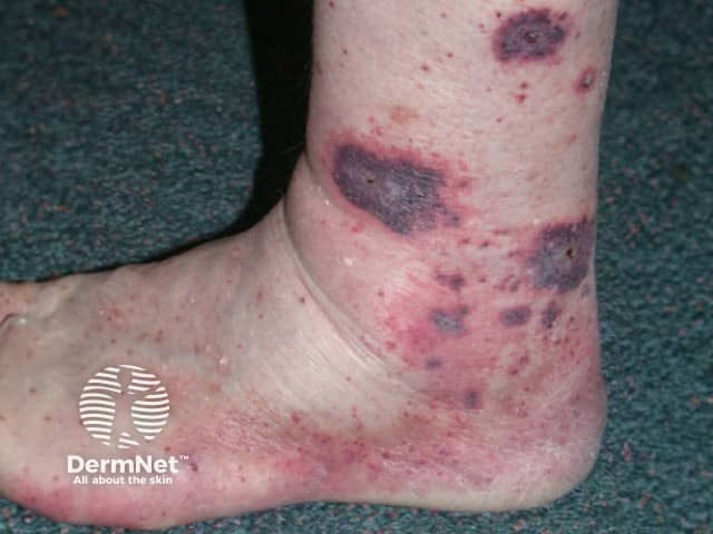
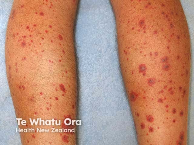
- Palpable purpura (purple, non-blanching papules and plaques)
- Sometimes, petechiae and ecchymoses
- Non-purpuric erythematous or urticaria-like weals, macules and papules
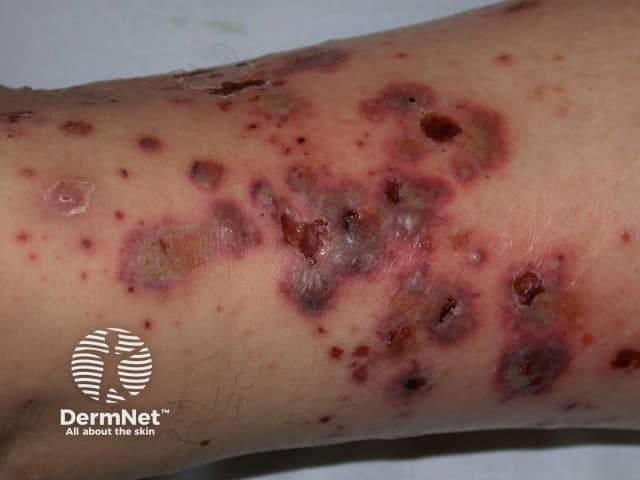
- Haemorrhagic bullae, necrosis and superficial ulceration
- Local pruritus, burning pain and swelling
- Variable systemic symptoms with fever, joint aches, lymphadenopathy and gastrointestinal upset.
The initial acute rash of small vessel vasculitis usually subsides within 2–3 weeks, but crops of lesions may recur over weeks to several months, and hypersensitivity vasculitis may rarely become relapsing or chronic.

Cutaneous vasculitis

Cutaneous vasculitis

Hypersensitivity vasculitis
Complications of small vessel vasculitis
The most severe complications of small vessel vasculitis are seen with its systemic forms, such as kidney disease associated with Henoch-Schönlein purpura.
Local ulceration from cutaneous small vessel vasculitis can lead to:
- Wound infection or cellulitis
- Chronic leg ulceration
- Scar formation.
How is cutaneous small vessel vasculitis diagnosed?
The clinical diagnosis of an acute cutaneous small vessel vasculitis is generally straightforward.
Thorough history and examination are essential to determine if symptoms and signs are confined to the skin, or if there may be systemic involvement, and to establish a cause.
Cutaneous small vessel vasculitis is confirmed by 4-mm punch biopsy of an early purpuric papule, ideally present for 24–48 hours. Histopathology reveals neutrophils around arterioles and venules, and fibrinoid necrosis (fibrin within or inside the vessel wall). There may be extravasated red cells, leukocytoclasis (broken-up neutrophils within the vessel wall) and signs of an underlying disease.
Direct immunofluorescence (DIF) of a lesion less than 24 hours old often reveals immunoglobulins and complement.
- Perivascular IgA is characteristic of Henoch-Schönlein purpura
- Perivascular IgM suggests cryoglobulinaemia or rheumatoid arthritis
- Interface immunoglobulins may be a sign of systemic lupus erythematosus
- DIF is often negative in cutaneous polyarteritis nodosa and ANCA-positive vasculitis
Screening tests are requested to identify any underlying cause and to determine the extent of involvement of internal organs. The history and symptoms may suggest these. Patients should have:
- Urinalysis, looking for protein, blood and glomerular casts
- Full blood count, liver and kidney function
Additional tests may include:
- Anti-nuclear antibodies (ANA), extractable nuclear antigen profile (ENA)
- Anti-streptococcal antibodies, human immunodeficiency virus (HIV), hepatitis B and C serology
- Serum complement levels
- Protein and immunoglobulin electrophoresis
- Cryoglobulins
- Antineutrophil cytoplasmic antibody (ANCA)
- Chest X-ray if symptoms suggest lung disease
If an initial screen indicates an abnormality or there is clinical suspicion of a more widespread vasculitic process, further investigations will be requested. The majority of patients presenting with palpable purpura have primary cutaneous small vessel vasculitis, and no underlying cause is found in spite of extensive investigations.
What is the treatment for cutaneous small vessel vasculitis?
In most patients presenting with the first episode of acute cutaneous small vessel vasculitis, general measures are all that is required to keep the patient comfortable until the rash spontaneously resolves.
- If an underlying cause is found, remove the trigger (for example, stop the drug) and treat associated disease(s)
- Rest — exercise often induces new lesions
- Apply compression and elevate the affected limb(s)
- Use simple analgesics and NSAIDs for pain
- Protect fragile skin from injury
- Apply emollients to relieve dryness and itch
- Dress ulcers
- Treat secondary bacterial infection
Severe, recurrent or chronic cutaneous small vessel vasculitis
Medications used to control cutaneous vasculitis have not been subjected to randomised trials. They are recommended in acute vasculitis when ulcerated and in symptomatic relapsing or chronic disease. They include:
- Corticosteroids: prednisone 0.5 mg/kg/day for 1–2 weeks, tapered over 3–6 weeks
- Colchicine: 0.5 or 0.6 mg bd
- Dapsone: 50–100 mg daily.
Other treatments reported to be helpful in at least some cases include:
- Hydroxychloroquine
- Minocycline
- Immune modulators: methotrexate, azathioprine, mycophenolate, ciclosporin
- Rituximab
- Intravenous immunoglobulin.
If cutaneous vasculitis is a manifestation of systemic vasculitis, treatment of the systemic disorder is required.
How can cutaneous small vessel vasculitis be prevented?
Flares of cutaneous small vessel vasculitis can be minimised by rest, compression and elevation of lower legs.
Once a drug is identified as the cause of small vessel vasculitis, the patient should avoid it lifelong. It is not usually possible to prevent other forms of vasculitis.
What is the outlook for small vessel vasculitis?
Idiopathic/primary cutaneous small vessel vasculitis is self-limiting. Most cases resolve within a period of weeks to months. When the cause is an underlying disease, the vasculitis may recur at variable intervals after the initial episode.
It is common for an orange-brown discolouration of the skin due to haemosiderin deposition to persist for weeks or months after the inflammatory disease has settled.
The prognosis of systemic vasculitis is dependent upon the severity of involvement of other organs. If vasculitis affects the kidneys, lungs or brain, it can be life-threatening.
References
- Gota C. Overview of cutaneous small vessel vasculitis. In: UpToDate, Post TW (Ed), UpToDate, Waltham, MA. (Accessed on January 23, 2016.)
- Fett N. Management of adults with idiopathic cutaneous small vessel vasculitis. In: UpToDate, Post TW (Ed), UpToDate, Waltham, MA. (Accessed on January 23, 2016.)
- Fett N. Evaluation of adults with cutaneous lesions of vasculitis. In: UpToDate, Post TW (Ed), UpToDate, Waltham, MA. (Accessed on January 23, 2016.)
- Phillipps J, Lama C, Strickley J, Musiek A. Infective endocarditis is the leading cause of infection-associated cutaneous vasculitis: A single academic center dermatology consultant experience. J Am Acad Dermatol. 2024;90(6):1262-1264. Journal
On DermNet
- Pathology of leukocytoclastic vasculitis
- Cutaneous vasculitis
- Vascular skin problems
- Purpura
- Allergies explained
- Direct immunofluorescence
- Skin signs of rheumatic disease
Other websites
- Hypersensitivity Vasculitis — Medscape Drugs & Diseases
- Leukocytoclastic Vasculitis — Medscape Drugs & Diseases
- Cutaneous vasculitis — British Association of Dermatologists
- The Vasculitis Foundation
