Main menu
Common skin conditions

NEWS
Join DermNet PRO
Read more
Quick links
Acute haemorrhagic oedema of infancy — extra information
Acute haemorrhagic oedema of infancy
Author: Dr Andre Schultz, Paediatric Registrar, Waikato Hospital, Hamilton, New Zealand, 2007. Updated by A/Prof Amanda Oakley, Dermatologist, Hamilton, New Zealand, January 2016.
Introduction
Demographics
Causes
Clinical features
Diagnosis
Treatment
Outcome
What is acute haemorrhagic oedema of infancy?
Acute haemorrhagic oedema (hemorrhagic edema with the American spelling) is a rare type of cutaneous small vessel vasculitis with a characteristic presentation in infants.
It consists of a clinical triad of:
- Large bruise-like lesions (purpura)
- Swelling (oedema)
- Fever
Acute haemorrhagic oedema of infancy was originally described by Snow in the USA in 1913. Finkelstein described it in Europe in 1938 and it has been recognised in the European literature under various terms since:
- Finkelstein disease
- Seidlmayer syndrome
- Infantile postinfectious iris-like purpura and oedema
- Purpura en cocarde avec oedema.
Who gets acute haemorrhagic oedema?
Acute haemorrhagic oedema generally develops in children between the ages of 4 months and 2 years of age.
What causes acute haemorrhagic oedema?
There is uncertainty whether acute haemorrhagic oedema of infancy is a mild variant of Henoch-Schoenlein purpura (HSP) that occurs in infancy or a distinct clinical entity. Clinically it is similar to, but milder than HSP. It occurs in a more restricted age range and has different skin lesions.
The cause of acute haemorrhagic oedema is unknown. It is an immune-mediated process, possibly an immune complex disorder. Immune complexes are made up of aggregates of antibodies and the particles that these antibodies are directed against.
Many possible triggers for this immune-mediated disease have been reported.
Infectious agents |
|
||
Medications |
|
||
Immunisations |
|||
What are the clinical features of acute haemorrhagic oedema?
Acute haemorrhagic oedema of infancy is frequently preceded by a prodromal illness such as a viral upper respiratory tract infection. The striking characteristic features appear as described in the table below; however, the child remains in good general health.
Rash |
|
||
Oedema |
|
||
Fever |
|
||
Internal organ involvement |
|
||
Acute haemorrhagic oedema of infancy
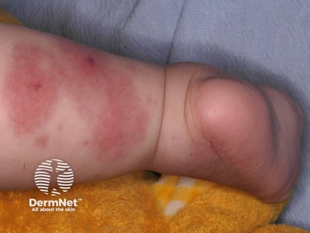
Finkelstein disease or acute haemorrhagic oedema of infancy
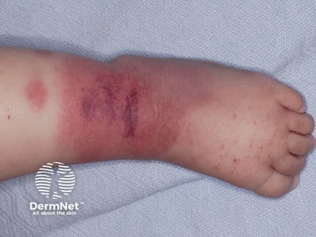
Finkelstein disease or acute haemorrhagic oedema of infancy
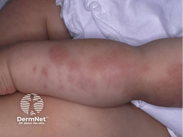
Finkelstein disease or acute haemorrhagic oedema of infancy
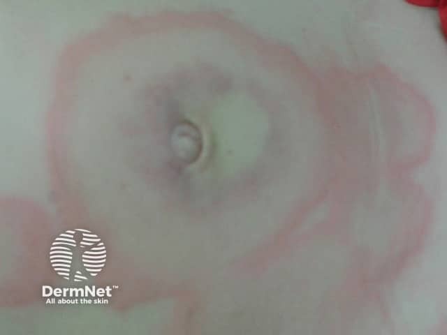
Finkelstein disease or acute haemorrhagic oedema of infancy
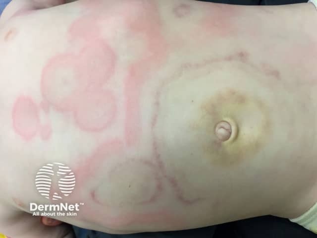
Finkelstein disease or acute haemorrhagic oedema of infancy
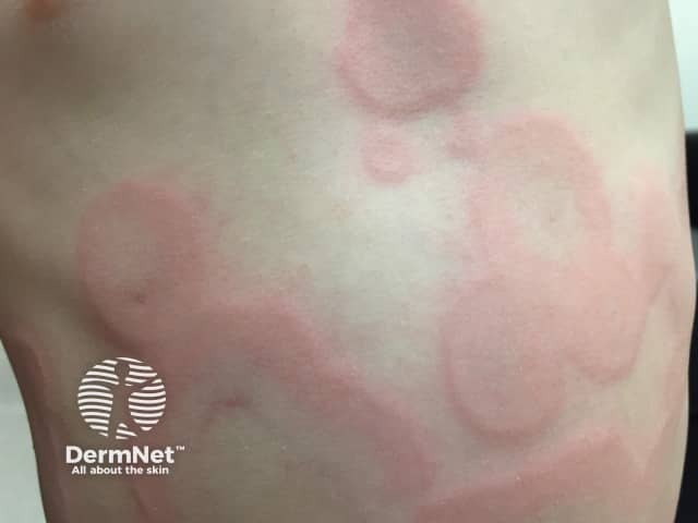
Finkelstein disease or acute haemorrhagic oedema of infancy
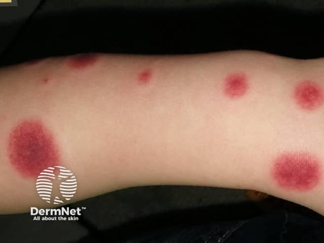
Acute haemorrhagic oedema
How is acute haemorrhagic oedema diagnosed?
Acute haemorrhagic oedema of infancy is usually diagnosed on clinical grounds alone. Other causes of purpura may first need to be excluded, as well as rashes that have a similar cockade pattern, like erythema multiforme, urticaria and Kawasaki disease. An inflicted injury should also be considered.
It is not usually necessary to perform skin biopsy if the diagnosis is thought of. Histopathology reveals a leukocytoclastic vasculitis (this means there are broken-up white cells involved with inflamed small blood vessels). Histopathologic findings are identical to HSP. However, the pattern of antibody staining on direct immunofluorescence of a skin biopsy is different; IgA is found in only one-third of patients with haemorrhagic oedema.
What is the treatment for acute haemorrhagic oedema?
No treatment is required as it resolves spontaneously. Systemic steroids and antihistamines do not alter the disease course.
What is the outcome for acute haemorrhagic oedema?
Acute haemorrhagic oedema of infancy usually resolves spontaneously over 1–3 weeks with complete recovery. Recurrence may occur but is uncommon, and usually occurs early.
References
- Infantile Henoch-Schonlein Purpura. Arch Fam Med. 2000; 9: 553–6.
- Acute Hemorrhagic Edema of Infancy. Dermatology Online Journal 2006; 12 (5): 10. Journal
- Acute Haemorrhagic Oedema of Childhood. Arch Dis Child. 2006; 91: 382.
On DermNet
Other websites
- Acute Hemorrhagic Edema of Infancy – Medscape Reference
