Main menu
Common skin conditions

NEWS
Join DermNet PRO
Read more
Quick links
Eruptive vellus hair cysts — extra information
Eruptive vellus hair cysts
Author: Dr Nick Turnbull, Dermatology Registrar, London, United Kingdom, 2011. Updated by Dr Amanda Oakley, 22 February 2014.
Introduction - vellus hair
Introduction - eruptive vellus hair cysts
Demographics
Causes
Clinical features
Other conditions
Diagnosis
Treatment
What is vellus hair?
Vellus hairs are fine, blond hairs normally growing on the face, trunk and limbs (in contrast to terminal hairs, which are longer, pigmented, and mainly arising on the scalp, beard, underarms and pubic area).
What are eruptive vellus hair cysts?
Eruptive vellus hair cysts are small papules containing vellus hairs. They are usually found on the central chest. Eruptive vellus hair cysts are quite rare.
Who gets eruptive vellus hair cysts?
Eruptive vellus hair cysts are likely to be familial when they occur in early childhood. Sporadic forms tend to develop later in teenage years. Boys and girls are equally affected.
They are usually an isolated finding and not associated with other skin abnormalities. Eruptive vellus hair cysts sometimes arise in children with anhidrotic and hidrotic ectodermal dysplasia, steatocystoma multiplex, or pachyonychia congenita.
What causes eruptive vellus hair cysts?
Eruptive vellus hair cysts are thought to be the result of occlusion (blockage) at the level of the infundibulum (the part of the hair follicle just below the epidermis), subsequent cystic dilatation (enlargement) of the hair follicle and secondary atrophy (withering) of the hair bulb. This is probably a developmental abnormality.
Keratin gene mutations are a likely association in familial cases when they have an autosomal dominant inheritance. This means an abnormal gene comes from one parent. In some cases, mutations have occurred in the gene that encodes keratin 17 (K17).
What are the clinical features of eruptive vellus hair cysts?
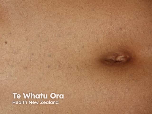
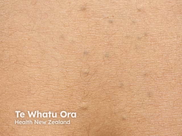
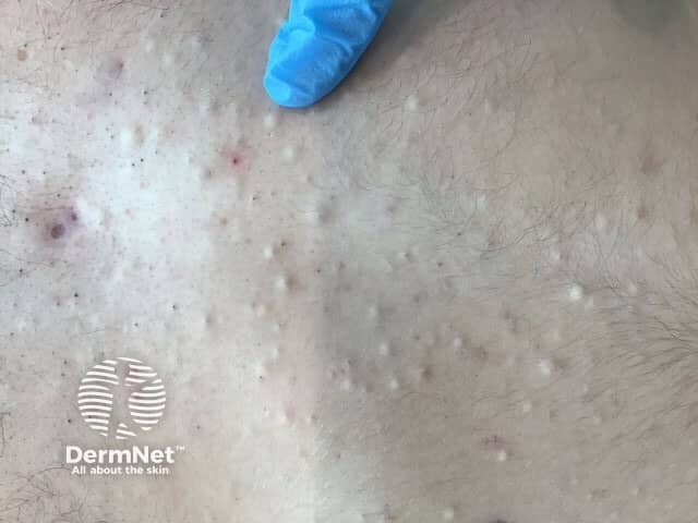
Vellus hair cysts usually present as small red or brown bumps over the sternum. They have also been reported to occur on the limbs and vulva. There may be few to numerous cysts, sometimes numbering in the hundreds.
Individual lesions are usually small smooth dome-shaped papules, 2–3 mm in size. They may be dimpled or umbilicated and sometimes have a scaly or crusty surface.
What other conditions should be considered?
Epidermoid/infundibular cysts and steatocystoma multiplex also present as asymptomatic papules or nodules on the anterior chest.
The differential diagnosis may include folliculitis and keratosis pilaris.
How are eruptive vellus cysts diagnosed?
The diagnosis of eruptive vellus hair cysts is often made clinically, because of the typical age of onset, the site of the lesions, and their appearance.
Incision or puncture of the cyst and examination of the contents under a microscope will reveal the vellus hairs.
A skin biopsy may confirm the diagnosis. Histopathology shows stratified-squamous epithelium with a granular layer that surrounds a cystic space filled with laminated keratin and a variable number of vellus hair shafts.
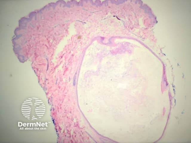
Low power view of cyst
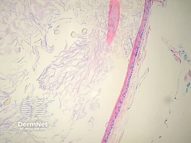
Hair shafts within cyst
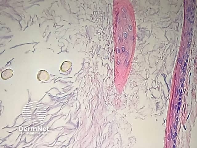
High power view
What is the treatment for eruptive vellus cysts?
In many cases, treatment is unnecessary as the lesions are harmless. The cysts disappear by themselves in about 25% of children.
Long term use of keratolytic creams containing urea or salicylic acid may be useful. Tretinoin and oral retinoids (isotretinoin, acitretin) have been used with limited success.
Individual lesions may be removed surgically by excision, curettage, cryotherapy or laser but this is likely to result in scarring.
References
- Weedon, D. 2008. Weedon's Skin Pathology. 2nd Edition. Elsevier.p429
- Bolognia JL, Jorizzo JL, Rapani RP. Dermatology: second edition. 2008: 1684-5
- Eruptive Vellus Hair Cysts. – Cory A Dunnick, MD; Chief Editor: Dirk M Elston, MD. Medscape Reference
- Khatu S, Vasani R, Amin S. Eruptive vellus hair cyst presenting as asymptomatic follicular papules on extremities. Indian Dermatol Online J. 2013 Jul;4(3):213-5. doi: 10.4103/2229-5178.115521. PubMed PMID: 23984238; PubMed Central PMCID: PMC3752480.
- Patel U, Terushkin V, Fischer M, Kamino H, Patel R. Eruptive vellus hair cysts. Dermatol Online J. 2012 Dec 15;18(12):7. PubMed PMID: 23286797.
- Torchia D, Vega J, Schachner LA. Eruptive vellus hair cysts: a systematic review. Am J Clin Dermatol. 2012 Feb 1;13(1):19-28. doi: 10.2165/11589050-000000000-00000. Review. PubMed PMID: 21958358.
On DermNet
