Main menu
Common skin conditions

NEWS
Join DermNet PRO
Read more
Quick links
52-year-old with expanding abdominal plaques
Last reviewed: August 2023
Author: Dr Shadi Habib, Dermatologist, Ibn al-Walid Hospital, Homs, Syria (2023). Reviewing dermatologist: Dr Ian Coulson
Edited by the DermNet content department


Background
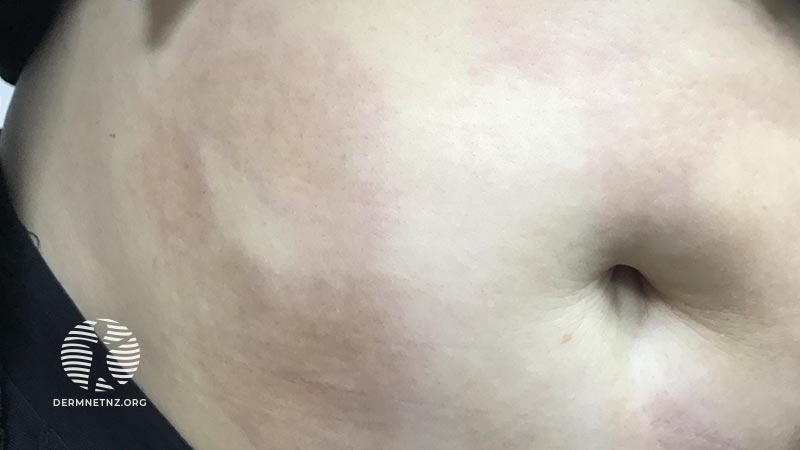
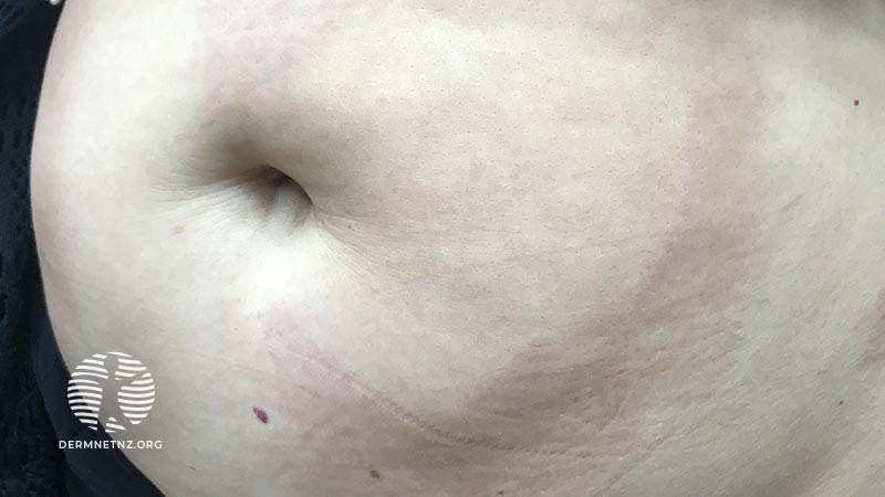
A 52-year-old female patient presented to our clinic complaining of red-brown asymptomatic multiple plaques with defined borders on the abdomen for one year with a peripheral expansion.
On examination, there were oval plaques on the abdomen with a sclerotic centre surrounded by a violaceous border, with some coalescing to form a larger plaque.
There was no mention of any systemic symptoms and laboratory tests were unremarkable.
What is the likely diagnosis?
A skin biopsy was obtained including subcutaneous fat, which demonstrated collagen bundles extending into the reticular dermis, enclosing the eccrine glands and blood vessels.
With the clinical and histological findings, the likely diagnosis is plaque morphoea (morphea).
There are many subtypes of morphoea such as plaque, linear, generalised, and pansclerotic. It is important to evaluate the patient for systemic symptoms and with laboratory tests such as ANA, anti-histone antibodies, and anti-SS DNA antibodies.
How should she be managed?
In patients with superficial circumscribed lesions, topical treatments are appropriate such as:
- Topical corticosteroids
- Topical tacrolimus 0.1%
- Imiquimod.
For patients with more widespread lesions and deep morphoea, consider:
- UVA1 phototherapy
- Narrowband UVB phototherapy.
Rapidly progressive lesions require treatment with systemic corticosteroids or methotrexate.
What is the prognosis?
Morphoea can be chronically active or relapsing and remitting. Deep tissue involvement (eg, subcutaneous tissue, muscle, or bone) can lead to deformity and dysfunction.
