Main menu
Common skin conditions

NEWS
Join DermNet PRO
Read more
Quick links
Syringofibroadenoma pathology — extra information
Syringofibroadenoma pathology
Author: Dr Ben Tallon, Dermatologist/Dermatopathologist, Tauranga, New Zealand, 2011.
Histology
Special stains
Histological variants
Differential diagnoses
Syringofibroadenoma is a benign adnexal tumour thought to be of eccrine origin, also named syringofibroadenoma of Mascaro, after the man who first described this tumour. It typically occurs on the extremities.
Histology of syringofibroadenoma
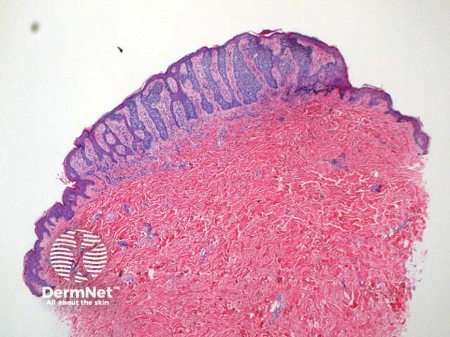
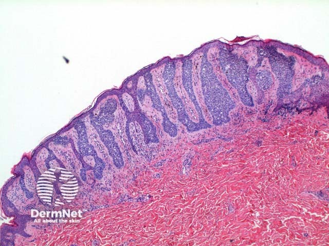
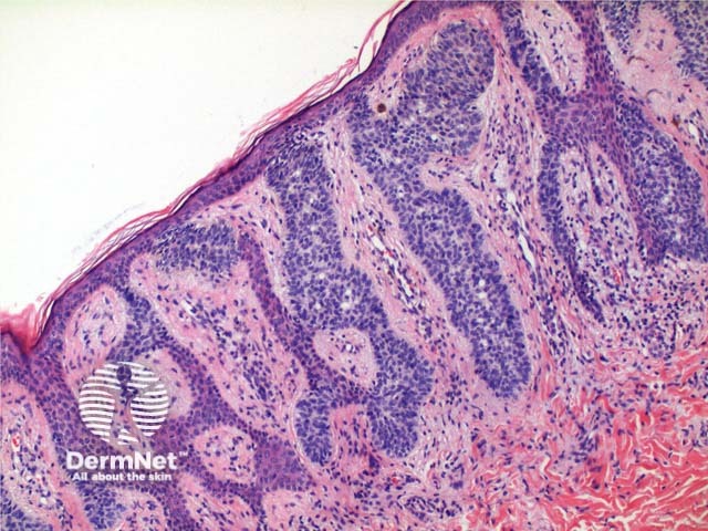
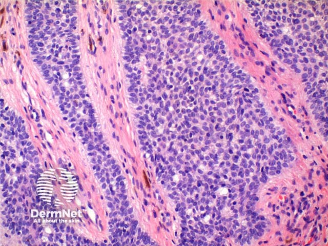
Scanning power view of the histology of syringofibroadenoma reveals an epidermal proliferative process (Figure 1). Vertically oriented anastamosing strands of basaloid epithelium are seen arising from multiple points along the epidermis (Figure 2). The basaloid cells are smaller than adjacent keratinocytes and show mild variation in size (Figure 4). Ductal differentiation is seen (Figures 3 and 4). The tumour has notable intervening fibrovascular stroma, and may have a mild superficial lymphocytic infiltrate (Figure 4).

Figure 1

Figure 2

Figure 3

Figure 4
Special stains in syringofibroadenoma
CEA (CarcinoEmbryonic Antigen) staining will identify ductal differentiation by highlighting the luminal cells.
Histological variants of syringofibroadenoma
A clear cell variant has been described, which shows cytoplasmic clearing.
Multiple syringofibroadenomas have been seen in association with the ectodermal dysplasias syndromes of Clouston and Schopf.
Reactive eccrine syringofibroadenoma demonstrates the same histological features but is seen adjacent to or in association with various inflammatory diseases.
Differential diagnosis of syringofibroadenoma
Basal cell carcinoma (fibroepithelioma type): The tumour strands will show focal changes typical of basal cell carcinoma with peripheral palisading and clefting artefact. There is a loose fibrous stroma.
Eccrine poroma: This tumour shows a more uniform small epithelial cell proliferation with vertical columns extending into the dermis.
Acrosyringeal naevus: Strong PAS positivity and a plasma cell infiltrate here are the reported distinguishing features.
Syringofibroadenocarcinoma: This is seen as an area of transformation within an existing syrigofibroadenoma where a malignant phenotype emerges displaying cytological atypia.
References
- Skin Pathology (2nd edition, 2002). Weedon D
- Pathology of the Skin (3rd edition, 2005). McKee PH, J. Calonje JE, Granter SR
