Main menu
Common skin conditions

NEWS
Join DermNet PRO
Read more
Quick links
Surgical treatment of stable vitiligo — extra information
Treatments Autoimmune/autoinflammatory
Surgical treatment of stable vitiligo
Author: Niranthari Chinniah MBBS, Dermatology Registrar, Sydney, Australia; Dr.Monisha Gupta, MD, FACD, Dermatologist, Sydney, Australia; Chief Editor: Dr Amanda Oakley, Dermatologist, Hamilton, New Zealand, July 2014.
Introduction
How it works
Different types
Grafting of melanocyte-rich tissue
Grafting of melanocyte cell suspensions
Contraindications and cautions
What is vitiligo?
Vitiligo is a chronic skin disorder that causes areas of skin to lose colour. It presents as depigmented (white) patches. Exposed body sites, such as the face, elbows, knees, hands and feet, are often involved, resulting in significant cosmetic concerns. Vitiligo is usually treated with creams and tablets, or by phototherapy. Vitiligo may fail to improve or clear with these treatments.
Surgical treatment options can be considered in patients with stable vitiligo.
What is the goal of vitiligo surgery?
The goal of vitiligo surgery is to achieve complete repigmentation that cosmetically matches the surrounding normal skin.
Appropriate patient selection is vital in ensuring the best outcome, as not all patients or vitiliginous skin sites are suitable for surgery. Prior to surgery the following patient factors should be considered:
- The age of the patient
- Patient expectations
- Disease stability
- Size and location of the vitiligo patch
- The proposed method of surgery
- The proposed donor site
What are the different types of vitiligo surgery?
All types of surgical treatment aim to transfer melanocytes (pigment-producing cells) from normal skin (the donor site) to the skin affected by vitiligo.
Surgical treatment for vitiligo can be considered in two main categories:
- Grafting of melanocyte-rich tissue (tissue grafting)
- Grafting of melanocyte cells (cellular grafting).
Commonly used surgical techniques for repigmentation surgery are listed below
Types of vitiligo surgery |
|
|---|---|
Grafting of melanocyte-rich tissue |
Grafting of melanocyte cell suspensions |
|
|
The superficial or uppermost layer of affected skin is usually removed under local anaesthesia in an outpatient setting. Techniques to remove the skin include:
- Dermabrasion
- Cryotherapy
- Shave biopsy
- Punch biopsy
- Laser therapy
Grafting of melanocyte-rich tissue
Miniature punch grafting
Miniature punch grafting is one of the most commonly used techniques, due to its simplicity and efficacy. Bits of skin about 2 mm in diameter are punched out from the donor site on buttock or thigh and placed on the donor site of vitiliginous skin, where recipient chambers have also been created by punches.
Potential immediate complications of miniature punch grafting include:
- Loss of graft tissue
- Infection
Mid- to long-term complications of miniature punch grafting include:
- Hyperpigmentation
- Imperfect colour matching
- Peripheral depigmentation (halo effect)
- Graft rejection
- Keloid and hypertrophic scar at the donor or graft site
- Cobblestone appearance (more common with larger punch biopsies)
- Persistent vitiligo
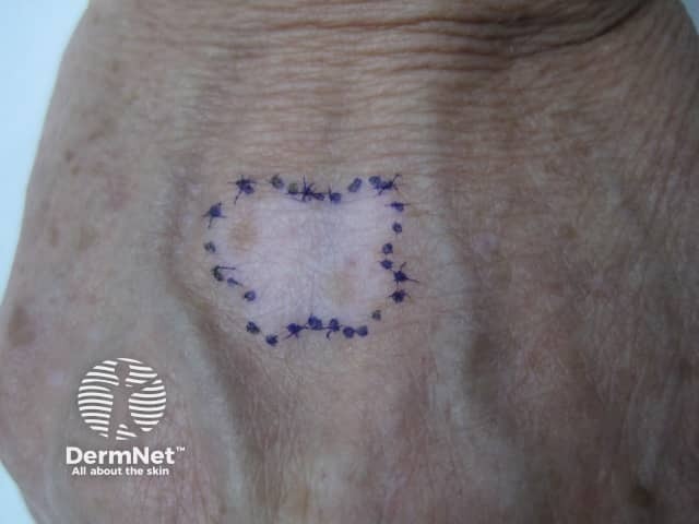
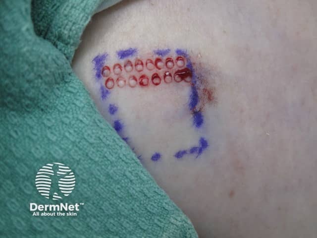
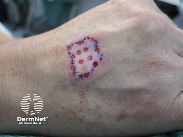
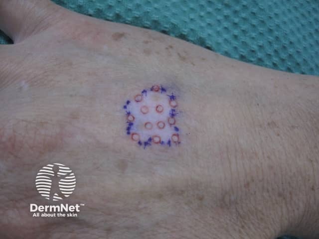
Punch grafting in vitiligo
Vitiligo surgery
Vitiligo surgery
Vitiligo surgery
Vitiligo surgery
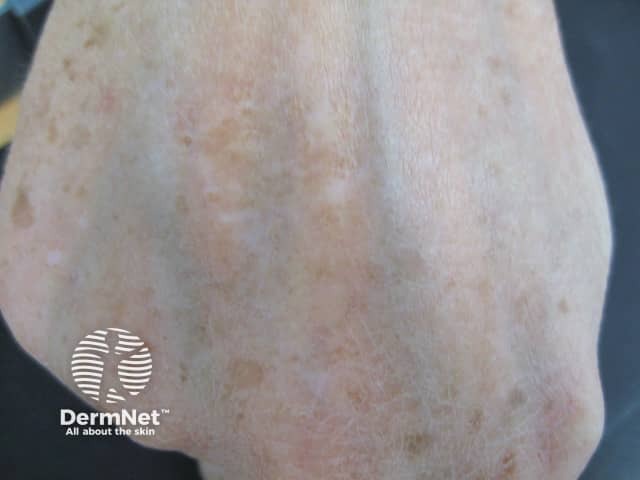
Vitiligo surgery
Recipient site, back of hand (a). Two-mm diameter full-thickness skin grafts are removed from a pigmented donor area on the thigh (b), using a punch (c). At the same time, equal sizes of holes are made in the white-skinned recipient area (d). The punched out pieces of skin from the donor area are then transplanted into the recipient area (e). About 30 grafts can be easily inserted at one sitting. The process may be repeated to treat larger areas. Outcome (f). Courtesy of Dr M.Gupta, Sydney, Australia.
Suction blister grafting
In suction blister grafting, negative pressure is applied to the normally pigmented donor site to promote the formation of multiple blisters.
Blisters may be raised using one of the following options:
- Syringe
- Suction pump
- Suction cups
- Negative pressure cutaneous suction chamber system
The bases of syringes of sizes 10 ml and 20 ml are coated with vaseline and are applied on the donor site. It usually takes 1.5 to 2.5 hours for the development of blisters. The roofs of the blisters (the grafts) are surgically removed, cut to the appropriate size and shape, and transplanted onto the prepared recipient site.
Good cosmetic results can be achieved, with minimal scarring of the donor site or cobblestoning at the recipient site. Suction blister grafting is generally safe, easy to perform and inexpensive, with good success rates. However, it can also be very time consuming, and can be performed only on small areas of skin.
Complications of suction blister grafting are limited to:
- Hyperpigmentation of the donor site
- Graft rejection
- Depigmentation around the graft.
Split thickness skin grafting
Split thickness skin grafting involves shaving off thin layers of skin from the donor site. In comparison to punch grafting and suction blister grafting, split thickness skin grafting can cover larger areas and produces uniform pigmentation with no cobblestoning.
Complications of split thickness skin grafting include:
- Hyperpigmentation
- Peripheral depigmentation
- Milia formation
- Graft rejection
Grafting of melanocyte cell suspensions
Autologous non-cultured epidermal cell suspensions
Autologous non-cultured epidermal cell suspensions are gaining popularity around the world as the treatment of choice for surgical management of vitiligo. Pre-prepared kits are available.
Tissue is harvested from a donor site and is incubated with trypsin to separate the epidermis from the dermis. The melanocytes are then separated from the epidermis and made into a cell suspension that can then be transplanted onto the de-epithelialized recipient skin.
Autologous non-cultured epidermal cell suspensions allow large areas to be treated in one session using a small donor graft. They result in excellent colour matching. However the procedure is expensive and complex. Due to stringent rules regarding tissue handling in Australia and elsewhere, it is not yet widely available.
Cultured melanocyte suspensions
With cultured melanocyte suspensions, tissue is also harvested and incubated with trypsin. However, after separation of the epidermis, the melanocytes and keratinocytes are incubated in a medium containing growth factors. The cultured suspension is then transplanted on to de-epithelialized recipient skin.
Cultured melanocyte suspensions allow a large area to be treated in a single session. It uses a larger donor-to-recipient ratio than the non-cultured technique. This sophisticated technique requires a special laboratory.
What are the contraindications and cautions of vitiligo surgery?
Vitiligo surgery is used in stable disease, ie vitiligo that does not progress over a period of 6–12 months. Segmental vitiligo generally has better surgical outcomes than non-segmental vitiligo. Outcome also depends on body site, with surgical treatment of genitals, lips, eyelids and bony prominences particularly variable.
Because the progress of the disease can be difficult to predict, vitiligo surgery is contraindicated in:
- Active unstable vitiligo.
- Childhood vitiligo
References
- Felsten LM, Alikhan A, Petronic-Rosic V; Vitiligo: A comprehensive overview. Part II: Treatment options and approach to treatment. J Am Acad Dermatol 2011; 65 (3):493–514. PubMed
- Falabella R; Surgical Approaches for Stable Vitiligo. Dermatol Surg 2005; 31:1277–84. PubMed
- Patel NS, Paghdal KV, Cohen GF; Advanced treatment modalities for vitiligo. Dermatol Surg 2012; 38:381–91. PubMed
- Khunger N, Kathuria SD, Ramesh V. Tissue grafts in vitiligo surgery – past, present, and future. Indian J Dermatol. 2009;54(2):150–8. PubMed
- Van Geel N, Ongenae K, De Mil M, Vander Haeghen Y, Vervaet C, Naeyaert JM. Double-blind Placebo-Controlled Study of Autologous Transplanted Epidermal Cell Suspensions for Repigmenting Vitiligo. Arch Dermatol 2004; 140(10):1203–8. PubMed
On DermNet
Other websites
- Surgical Therapies for Vitiligo — Vitiligo Support International
- Surgery; Guidelines for the Management of Vitiligo. The European Dermatology Forum Consensus — Medscape News 2013
