Main menu
Common skin conditions

NEWS
Join DermNet PRO
Read more
Quick links
Spindle cell melanoma — extra information
Spindle cell melanoma
Author: Dr Daniel Yiu, Foundation Year Doctor, Department of Infectious Diseases, Wexham Park Hospital, Frimley Health NHS Trust, Slough, Berkshire, UK. DermNet Editor in Chief: Adjunct A/Prof. Amanda Oakley, Dermatologist, Hamilton, New Zealand. Copy edited by Gus Mitchell/Maria McGivern. January 2019.
Introduction
Demographics
Causes
Clinical features
Complications
Diagnosis
Differential diagnoses
Treatment
Outcome
What is spindle cell melanoma?
Spindle cell melanoma is a rare histological variant of melanoma, characterised by the presence of spindle-shaped melanocytes [1]. On microscopy, it is often mistaken for other skin and soft tissue cancers with spindle cell morphologies.
Desmoplastic melanoma is a variant of spindle cell melanoma where there are varying proportions of spindle cells and desmoplastic cells present in the histology.
Further studies are needed to clarify the clinical manifestations, risk factors, treatment management, and prognosis of spindle cell melanoma.
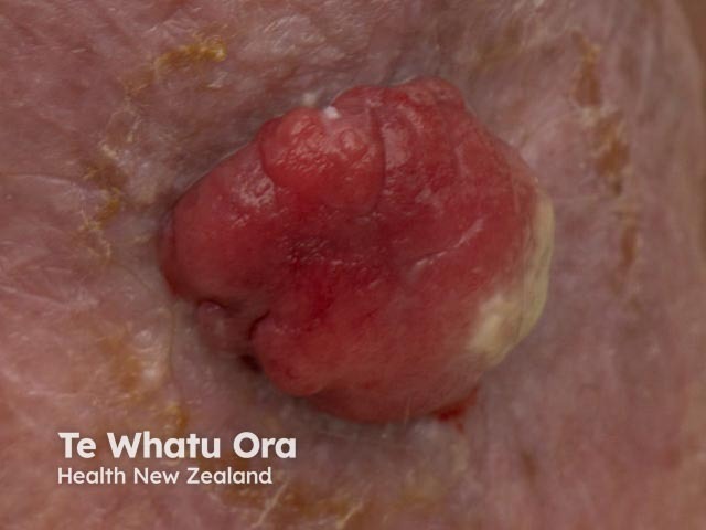
Spindle cell melanoma
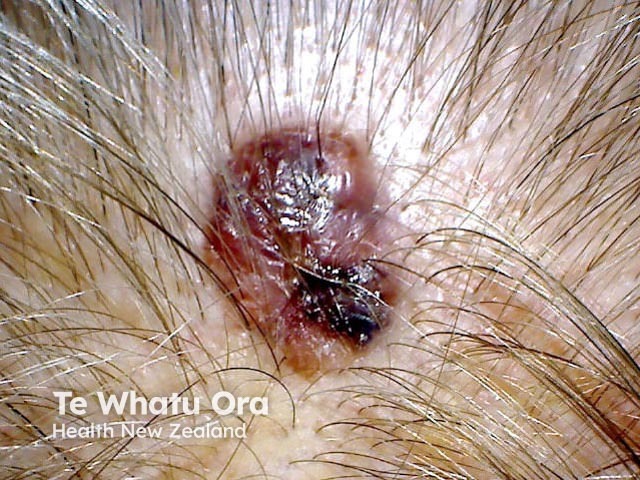
Spindle cell melanoma
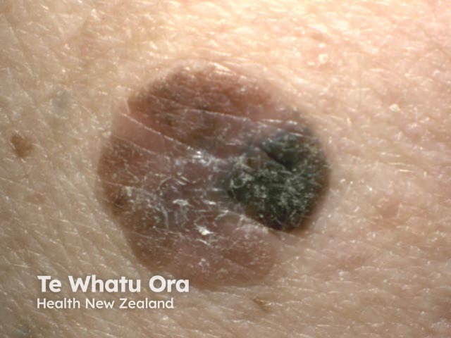
Spindle cell melanoma
Who gets spindle cell melanoma?
The actual incidence of spindle cell melanoma is unknown. Studies have suggested that between 1% and 14% of melanomas are of the spindle cell variant (including desmoplastic melanoma) [2,3].
Spindle cell melanomas more commonly occur in Caucasian men, affecting men and women at a ratio of 1.6:1–1.9:1 respectively. The average age at diagnosis is 50–80 years [1,4].
What causes spindle cell melanoma?
The exact cause of spindle cell melanoma is unknown. Spindle cell melanoma shares some common mutations with conventional (epithelioid) melanoma.
- Approximately 30% of spindle cell melanomas contain BRAF mutations; the V600E substitution mutation is the most common.
- NRAS and KIT mutations are rarely seen [4].
An association between spindle cell melanoma (including desmoplastic melanoma) and lentigo maligna has been observed in sun-exposed areas [5].
What are the clinical features of spindle cell melanoma?
Spindle cell melanoma frequently presents as a non-specific amelanotic (non-pigmented) nodule on the patient's trunk, head, or neck [1,3].
It may also first present as widespread melanoma metastases.
What are the complications of spindle cell melanoma?
Metastasis is the major complication of spindle cell melanoma. Delays in diagnosis can occur due to its atypical presentation histologically [1–3].
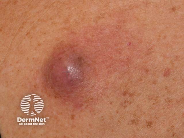
Spindle cell melanoma
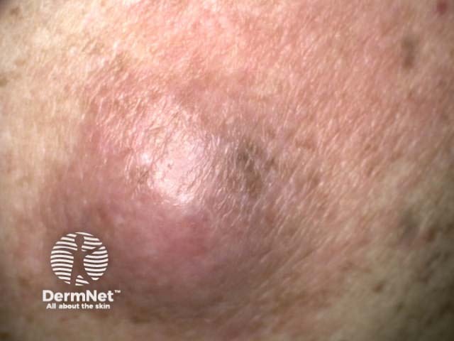
Spindle cell melanoma
How is spindle cell melanoma diagnosed?
Its non-specific features may lead to delays in the diagnosis of spindle cell melanoma, which is often not suspected clinically. The diagnosis is generally made on a biopsy of the lesion, but it can also be commonly mistaken for another tumour histologically [6].
A combination of histological clues and immunohistochemistry markers are required to diagnose spindle cell melanoma [3].
Features of spindle cell melanoma under microscopy include:
- An abundance of spindle-shaped tumour cells (> 90% of a tumour)
- Uniform, wavy, and slender nuclei, with variable size and shapes (pleomorphism) and variable nuclear atypia (abnormal appearance of cell nuclei)
- A high mitotic index (the ratio of cells undergoing mitosis over the total amount of cells)
- Greater cellular cohesion is seen on cytology than with epithelioid types of melanoma [2,7]
- A pagetoid spread of atypical melanocytes in spindle cell melanoma, associated with lentigo maligna [8].
Special stains may be used to differentiate spindle cell melanoma from other spindle cell tumours.
- The markers S100, SOX10, p75, HMB-45, laminin T, laminin NT, Melan-A, and c-KIT are positive after staining in spindle cell melanoma [2,3].
- In spindle cell melanoma, a higher proportion of cells show Ki-67, cyclin D1, and survivin after staining [9].
- Cytokeratin stains are negative in spindle cell melanoma [10,11].
What is the differential diagnosis for spindle cell melanoma?
Spindle cell melanoma can be easily confused with other spindle cell tumours. Others tumours it can be confused with are described below.
Desmoplastic melanoma
Desmoplastic melanoma was described as a variant of spindle cell melanoma in 1971 [6]. Recent studies now suggest that desmoplastic melanoma and spindle cell melanoma represent two distinct types of melanoma, as differences in staining, genetic mutations, and clinical manifestations have been found [3].
- When spindle cells represent > 90% of a tumour, a diagnosis of spindle cell melanoma is made.
- When > 90% of a tumour is composed of desmoplastic cells, a diagnosis of desmoplastic melanoma is made.
- Melanomas with both spindle cell and desmoplastic components at varying levels between 10% and 90% are referred to as mixed variants [2,3,12].
- Collagen IV, CD68, MDM2, and trichrome are positive after staining in desmoplastic melanoma [3].
- HMB-45, laminin T, laminin NT, Melan-A, and c-KIT are negative in desmoplastic melanoma [2,3].
Reed naevus
Reed naevus (also called a pigmented spindle-cell naevus) is a melanocytic naevus with a largely spindle-cell appearance under microscopy. The architecture is symmetrical with good lateral demarcation, epidermal hyperplasia, and uniform nests of cells [9].
Cutaneous clear-cell sarcoma
The differentiation of cutaneous clear cell sarcoma from spindle cell melanoma is largely histological, as both may stain positively for the same indicators: S100, HMB-45, and Melan-A. Cutaneous clear-cell sarcoma tends to show uniform patterns of spindle-cell fascicles (bundles) under microscopy, which are present throughout an entire tumour and encased by fibrous septa. The stroma tends to be hyalinised, sclerotic, and reticulated [13].
Leiomyosarcoma
Leiomyosarcoma pathology may be indistinguishable morphologically from melanoma.
- Smooth-muscle actin (SMA) stain is positive in leiomyosarcoma.
- The markers S100, p75, or HMB-45 are negative after staining [10,14].
Atypical fibroxanthoma
The markers S100, p75, and HMB-45 are negative after staining in atypical fibroxanthoma [10].
Fibrosarcoma
- Vimentin is positive in fibrosarcoma.
- The markers S100 and HMB-45 are negative after staining [5,14].
Peripheral nerve sheath tumours
- The markers S100, CD34, and GAP43 are positive after staining in peripheral nerve sheath tumours.
- HMB-45 and Melan-A are negative in peripheral nerve sheath tumours [10,15].
Dermatofibrosarcoma protuberans
- CD34 is positive in dermatofibrosarcoma protuberans.
- p75 is variable.
- S100 is negative [5,16].
Spindle-cell squamous cell carcinoma
- Cytokeratin stains such as 34betaE12 are positive in spindle-cell squamous cell carcinoma.
- S100 and HMB-45 are usually negative after staining.
- p75 is variable [10,11].
What is the treatment for spindle cell melanoma?
The treatment for spindle cell melanoma is similar to that for other forms of melanoma. Surgical excision is the first step in management [1].
What is the outcome for spindle cell melanoma?
Although the tendency for nodal involvement is low, the majority of spindle cell melanomas present with advanced disease, with a worse prognosis being seen in Caucasian men aged over 66 years [3].
Advanced disease with nodal and distal metastasis is associated with worse outcomes in spindle cell melanoma, as are higher grades of tumours, which have been associated with poorer disease-specific survival and overall survival [1].
References
- Xu Z, Shi P, Yibulayin F, Feng L, Zhang H, Wushou A. Spindle cell melanoma: incidence and survival, 1973–2017. Oncol Lett 2018; 16(4): 5091–9. PubMed
- Piao Y, Guo M, Gong Y. Diagnostic challenges of metastatic spindle cell melanoma on fine-needle aspiration specimens. Cancer 2008; 114(2): 94–101. PubMed
- Weissinger SE, Keil P, Silvers DN, Klaus BM, Moller P, Horst BA, et al. A diagnostic algorithm to distinguish desmoplastic from spindle cell melanoma. Mod Pathol 2014; 27(4): 524–34. PubMed
- Kim J, Lazar AJ, Davies MA, Homsi J, Papadopoulos NE, Hwu WJ, et al. BRAF, NRAS and KIT sequencing analysis of spindle cell melanoma. J Cutan Pathol 2012; 39(9): 821–5. PubMed
- Massi G, LeBoit PE. Histological Diagnosis of Nevi and Melanoma: Springer Berlin Heidelberg; 2013.
- Conlev J, Lattes R, Orr W. Desmoplastic malignant melanoma (a rare variant of spindle cell melanoma). Cancer 1971; 28(4): 914–36. PubMed
- Walia R, Jain D, Mathur SR, Iyer VK. Spindle cell melanoma: a comparison of the cytomorphological features with the epithelioid variant. Acta Cytol 2013; 57(6): 557–61. PubMed
- Barnhill RL, Crowson AN. Textbook of Dermatopathology: McGraw-Hill, Medical Pub. Division; 2004.
- Diaz A, Valera A, Carrera C, Hakim S, Aguilera P, Garcia A, et al. Pigmented spindle cell nevus: clues for differentiating it from spindle cell malignant melanoma. A comprehensive survey including clinicopathologic, immunohistochemical, and FISH studies. Am J Surg Pathol. 2011; 35(11): 1733–42. PubMed
- Sigal AC, Keenan M, Lazova R. P75 nerve growth factor receptor as a useful marker to distinguish spindle cell melanoma from other spindle cell neoplasms of sun-damaged skin. Am J Dermatopathol 2012; 34(2): 145–50. PubMed
- Morgan MB, Purohit C, Anglin TR. Immunohistochemical distinction of cutaneous spindle cell carcinoma. Am J Dermatopathol 2008; 30(3): 228–32. PubMed
- Miller DD, Emley A, Yang S, Richards JE, Lee JE, Deng A, et al. Mixed versus pure variants of desmoplastic melanoma: a genetic and immunohistochemical appraisal. Mod Pathol 2012; 25(4): 505–15. PubMed
- Hantschke M, Mentzel T, Rutten A, Palmedo G, Calonje E, Lazar AJ, et al. Cutaneous clear cell sarcoma: a clinicopathologic, immunohistochemical, and molecular analysis of 12 cases emphasizing its distinction from dermal melanoma. Am J Surg Pathol 2010; 34(2): 216–22. PubMed
- S, Metgud R, Naik S, Patel S. Spindle cell lesions: A review on immunohistochemical markers. Journal of Cancer Research and Therapeutics 2017; 13(3): 412–8. PubMed
- Chen WS, Chen PL, Lu D, Lind AC, Dehner LP. Growth-associated protein 43 in differentiating peripheral nerve sheath tumors from other non-neural spindle cell neoplasms. Mod Pathol 2014; 27(2): 184–93. PubMed
- West KL, Cardona DM, Su Z, Puri PK. Immunohistochemical markers in fibrohistiocytic lesions: factor XIIIa, CD34, S-100 and p75. Am J Dermatopathol 2014; 36(5): 414–9. PubMed
On DermNet
Other websites
- Spindle Cell Melanoma — NCI Thesaurus. National Institutes of Health. U.S. Department of Health and Human Services [cited November 17 2018].
- Melanoma risk assessment tool — Melanoma Institute of Australia
- Melanoma education — Melanoma Institute of Australia
