Main menu
Common skin conditions

NEWS
Join DermNet PRO
Read more
Quick links
Spider bite — extra information
Introduction
Spider species that bite
Signs and symptoms
Clinical features
Bacterial infections from spider bites
Treatment
Introduction
Spiders belong to the class of mainly terrestrial arthropods known as Arachnida. Medically significant classes of arachnids include spiders, ticks/mites and scorpions. Unfortunately through myths, legends and nowadays media, spiders have gained a reputation of being dangerous and harmful, and in some people instil a psychological fear known as arachnophobia. In reality, very few are dangerous to man and media reports exaggerating the dangers of spider bites are far out of proportion to the actual threat they pose.
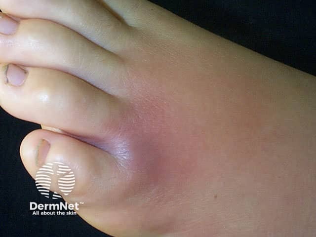
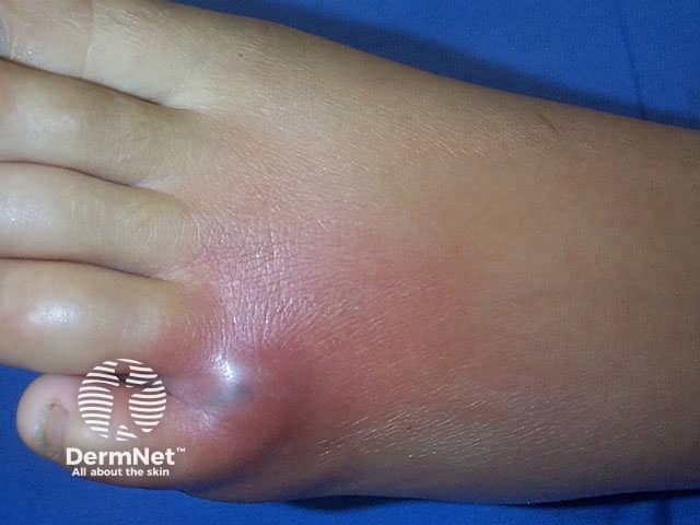
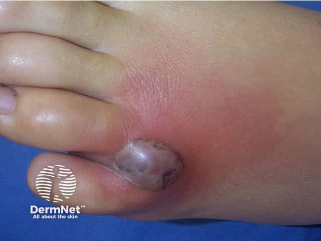
Necrotic spider bite

Day 1

Day 2

Day 3
Which spiders bite and may be harmful to man?
Latrodectus spp (widow spiders) are found throughout the world and known by many different common names according to country.
- Black widow (North America)
- Katipo (New Zealand)
- Red-back (Australia)
- Shoe-button (South Africa)
Loxosceles spp is found in South America, United States, Australia, and commonly in the tropics.
- Violin spiders
- Recluse spiders
- Brown recluse spiders
- Fiddleback spiders
Tegenaria agrestis (hobo spider) and Cheiracanthium (yellow sac spider) are found in the United States.
Phoneutria (banana spider) is found in Central and South America.
Atrax and Hadronyche (funnel-web spider) are found in Australia.
The venom produced by spider bites is generally either neurotoxic or cytotoxic. Web dwellers tend to have neurotoxic venom and non-web dwellers cytotoxic venom. Spiders of the Latrodectus genus produce neurotoxic venom, while the violin spider and yellow sac spider produce cytotoxic venom.
What are the signs and symptoms of a spider bite?
The signs and symptoms of a spider bite depend on many factors, these include:
- Neurotoxic or cytotoxic venom
- Amount of venom injected
- The health of the patient (e.g. any allergies)
- Age of the patient (small children and older persons are more adversely affected)
- Site of the bite.
The signs and symptoms from a bite from a spider with neurotoxic venom differ to those produced by a spider with cytotoxic venom. The severity of the symptoms depends on the species of the spider as the symptoms of bites from different species of Loxosceles can range from unremarkable (requiring no care), localised (usually self-healing), dermonecrotic (slow-healing ulcerated lesion requiring treatment), to systemic (vascular, renal damage and sometimes life-threatening).
Features of neurotoxic venom bite
- Affects neuromuscular junctions
- Severe pain in the chest and abdomen (cramp-like pains)
- Breathing difficulties, heart palpitations
- Nausea and vomiting
- Sweating, fever, excessive salivation
- Increased blood pressure
- Rash may develop
- Symptoms usually start about 1-3 hours after being bitten
- More severely affected are children and older persons
Features of cytotoxic venom bite
- Affects cellular tissue and usually restricted to the area of the bite
- Initial bite is painless but symptoms develop about 2–8 hours later, the area becomes painful and swollen
- Eventually, a blister may form over a necrotic lesion which then sloughs to create an ulcerated wound (up to 10cm)
- The ulcer will heal over months and leave behind a scar. In extreme cases, skin grafts may be necessary.
- In severe cases, systemic conditions may occur, such as thrombocytopenia, disseminated intravascular coagulation, renal failure
What are the dermatological features of a spider bite?
Widow spider bite
The skin around the site of the bite is red and two fang marks may be visible where the skin was penetrated. In untreated cases, a rash may develop after several days. Systemic symptoms are of more diagnostic value.
Violin or recluse spider bite
The dermatological features of these spider bites depend on the severity of the bite. In self-healing wounds, the bite site gets no worse than being swollen and red. With more serious bites a “bull's eye” wound may form. This is characterised by a central red swollen blister that is separated from a peripheral bluish region by a white zone of firm swelling. If the bite turns a purplish colour within the first few hours, this usually indicates severe localised tissue death (necrosis) may occur. Over days the blister forms a scab, which hardens and falls off to leave behind an ulcerated depression. Healing can take weeks to months.
Interestingly, it appears that bites that become systemic do not also develop necrotic wounds. It is thought that in necrotic wounds the venom is localised in the tissue whereas in systemic reactions the venom is distributed quickly throughout the body without any localised effects.
Another spider bite
- Hobo spider results in dermonecrotic lesions similar to violin/recluse spider bites.
- Yellow sac causes a painful, red, swollen and itchy bite that may produce a slightly necrotic wound that heals without scarring.
- Banana spider has few dermatological features, mainly neurotoxic symptoms.
- Funnel-web causes sudden onset of neurotoxic symptoms, and the dermatological features insignificant in comparison.
- Whitetail spider may cause discomfort and redness but does not cause ulceration or any serious symptoms.
Do spider bites cause bacterial infection?
Although many people attribute an episode of bacterial infection (especially cellulitis and necrotising fasciitis) to an unseen spider bite, they are falsely blamed. Documented spider bites have not led to skin these infections.
What is the treatment for a spider bite?
One of the most important aspects in treating spider bites it to try and identify the offending spider. The venom of spider bites is quite variable hence identification of the spider can be of value in determining the management of the condition.
General measures that should occur after a spider bite include:
- Wash the area well with soap and water
- Apply a cold flannel or ice pack wrapped in a cloth to the site
- Give paracetamol for pain
- Seek immediate emergency care for further treatment.
Depending on the identification of the offending spider and the severity of the bite, treatment may include:
- Muscle relaxants
- Stronger pain relievers
- Antihistamines to reduce swelling
- Antibiotics for confirmed secondary infection
- Supportive care
- Antivenin.
Specific treatment for bites from certain spiders include:
- Antivenin is available for bites by spiders of the Latrodectus and Loxosceles genera and is very effective if given soon after the bite.
- Hydroxyzine (antihistamine) may be given to alter the necrotic lesion of bites from spiders of the Loxosceles genus.
- Intravenous calcium gluconate alternating with methocarbamol to relieve muscle cramps caused by spiders belonging to the Latrodectus genus (e.g. black widow) (controversial).
- It is possible that systemic steroids may be of benefit.
References
- Book: Textbook of Dermatology. Ed Rook A, Wilkinson DS, Ebling FJB, Champion RH, Burton JL. Fourth edition. Blackwell Scientific Publications.
- Vetter RS, Visscher PK. Bites and stings of medically important venomous arthropods. Int J Dermatol. 1998 Jul;37(7):481–96. Review. PubMed PMID: 9679688.
- Study Rules Out Spiders as Common Cause of Bacterial Infections in Humans — UCR today, January 2015
On DermNet
- Erosions and ulcers
- Insect bites and stings
- Bee and wasp stings
- Arthropod infestations online course for health professionals
Other websites
- Spider Research — University of California Riverside
- Tick Bites — Medline Plus
- Spider Bites — Medscape Reference
- Brown Recluse Spider Bite — emedicinehealth
- White-tailed spiders — Landcare Research NZ
