

Palisaded and encapsulated neuroma pathology — extra information
Introduction Histology Special studies Differential diagnoses
Introduction
Palisaded and encapsulated neuroma is otherwise known as a solitary circumscribed neuroma. These lesions are typically small dermal nodules that occur on the face.
Histology of palisaded and encapsulated neuroma
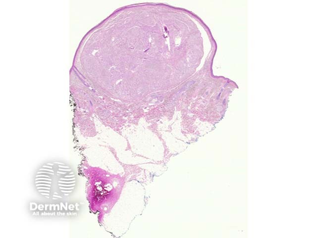
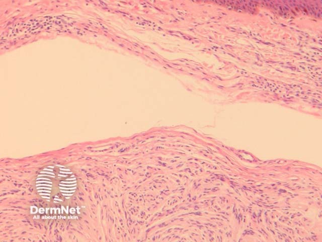
Palisaded and encapsulated neuroma is a circumscribed dermal tumour underlying an uninterrupted or attenuated epidermis (figure 1). A split between the tumour and the surrounding dermis is often seen (figures 1, 2).
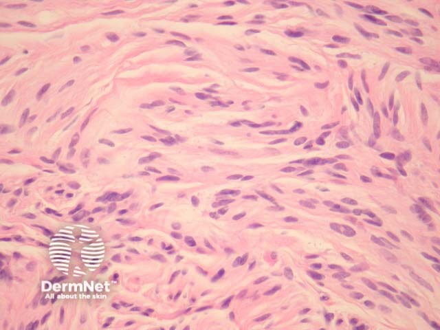
The tumour is composed of a loose matrix and fascicles of spindled cells, which resemble schwannoma (figure 3).
Special studies for palisaded and encapsulated neuroma
None are generally needed. The lesion is S100 positive. The surrounding capsule exhibits EMA positivity. Axons course through the lesion and can be demonstrated with neurofilament.
Differential diagnosis of a palisaded and encapsulated neuroma
Schwannoma – Its relationship with schwannoma has been questioned. Schwannomas display a varied cell density (Antoni A and B areas) and lack axons which are thought to course through palisaded and encapsulated neuroma.
Neurofibroma – These are typically non-encapsulated (with the exception of plexiform neurofibroma).
References
- Weedon’s Skin Pathology (Third edition, 2010). David Weedon
On DermNet