Main menu
Common skin conditions

NEWS
Join DermNet PRO
Read more
Quick links
Osteoma cutis pathology — extra information
Lesions (benign) Diagnosis and testing
Osteoma cutis pathology
Author: Dr Ben Tallon, Dermatologist/Dermatopathologist, Tauranga, New Zealand, 2010.
The reasons for bone formation in the skin are numerous, but can broadly be divided into primary type of osteoma cutis where no pre-existing disorder exists, or secondary. From a pathology perspective differentation between these two categories is all that is required, as clinical features determine the further breakdown within the primary group. Identification of the underlying process will form the basis for classifying bone deposition as secondary.
Histology of osteoma cutis
Scanning power of osteoma cutis reveals the presence of dense eosinophilic deposits in the dermis or subcutaneous tissue (Figure 1). Spicules of bone may be seen to perforate the epidermis in the process of transepidermal elimination (Figures 2 and 3). Most cutaneous bone formation occurs by the process of membranous ossification and so associated cartilage tissue is lacking. Bone is identified by osteocytes held within small lacunae (Figures 4 and 5) and the hydroxyapatite eosinophilic support material. In larger deposits, Haversian systems can be seen as concentric osteocytes around a central blood vessel (carried within a Haversian canal).
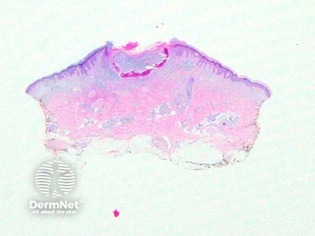
Figure 1
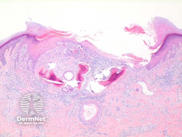
Figure 2
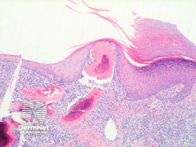
Figure 3
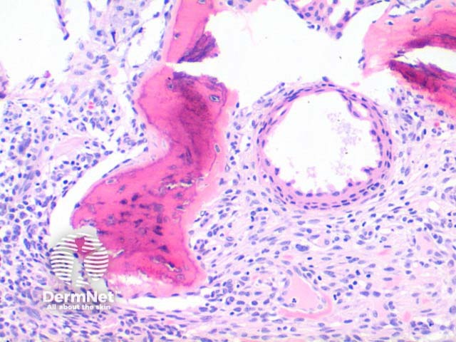
Figure 4
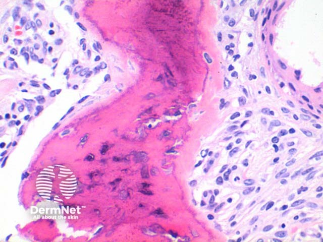
Figure 5
Histological variants of osteoma cutis
In secondary osteoma cutis underlying conditions include numerous inflammatory conditions and tumours such as basal cell carcinoma, cellular blue nevus, acne, morphoea profunda and pseudoxanthoma elasticum.
References
- Skin Pathology (2nd edition, 2002). Weedon D
- Pathology of the Skin (3rd edition, 2005). McKee PH, J. Calonje JE, Granter SR
On DermNet
