Main menu
Common skin conditions

NEWS
Join DermNet PRO
Read more
Quick links
Linear focal elastosis — extra information
Linear focal elastosis
Authors: Dr Beth Wright, Medical Registrar, Perth, Australia, 2012. Updated by Dr Julia Zhu, Dermatology Registrar and Dr Caroline Mahon, Dermatologist, Christchurch, New Zealand. DermNet Editor in Chief: Adjunct A/Prof. Amanda Oakley, Dermatologist, Hamilton, New Zealand. Copy edited by Gus Mitchell. January 2020.
Introduction Demographics Causes Clinical features Differential diagnoses Diagnosis Treatment
What is linear focal elastosis?
Elastosis refers to abnormal or increased deposition of elastin fibres within the dermis.
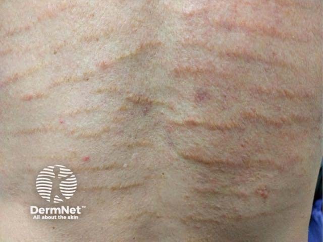
Linear focal elastosis is an uncommon form of dermal elastosis that resembles stretch marks.

Who gets linear focal elastosis?
There are few case reports of linear focal elastosis partly because it can be mistaken for more common stretch marks. The true prevalence of focal elastosis is unknown [1].
More cases of linear focal elastosis have been reported in males than in females. The average age range of onset is adolescence and ranges between 7 and 89 years [1,2]. There is no racial or ethnic predilection [3]. Strong hereditary factors have not been identified [1].
What causes linear focal elastosis?
The pathogenesis of linear focal elastosis is uncertain.
Linear focal elastosis and striae distensae may co-exist in the same distribution in affected individuals [3]. Linear focal elastosis may represent excessive regeneration of damaged elastic fibres as part of the repair process of striae distensae [6]. However, the clinical and histopathological appearances of linear focal elastosis and striae distensae differ.
No clear trigger factor has been identified for linear elastosis although blunt trauma, pregnancy, weight loss, or rapid growth may be involved [1].
What are the clinical features of linear focal elastosis?
Linear focal elastosis presents as yellow, palpable lines extending horizontally over the lower back. There are also reports of linear focal elastosis on the trunk, lower limbs and face [4,5].
Linear focal elastosis is usually asymptomatic and is diagnosed incidentally. It is not known to be associated with systemic illness.
What is the differential diagnosis for linear focal elastosis?
The main differential diagnosis for linear focal elastosis is striae distensae. However, striae are usually red or white and are depressed on palpation.
How is linear focal elastosis diagnosed?
Linear focal elastosis is diagnosed clinically by its appearance, distribution, and skin biopsy.
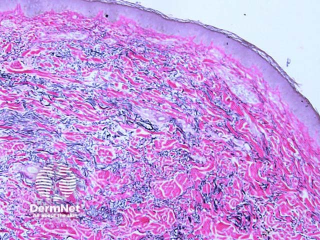
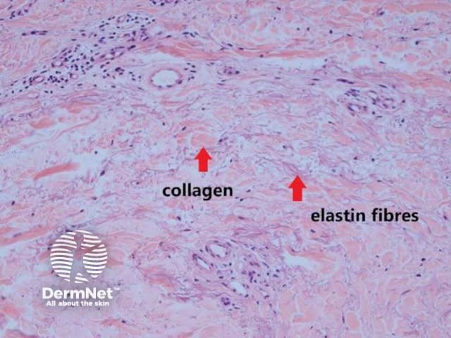
Histology demonstrates increased elastic fibres within the mid dermis. The fibres present as elongated material separating the dermal collagen. Elastic tissue stain can be useful to appreciate the elastin excess. Early lesions may paradoxically show elastolysis (destruction of elastin fibres) [1].


How is linear focal elastosis treated?
No effective treatment for linear focal elastosis has been described. Topical steroid and topical retinoid have shown no benefit [1].
References
- Seol JE, Kim DH, Cho GJ, Park SH, Jung SY, Kim H. Linear focal elastosis: a case report and institutional case series of 22 patients. Australas J Dermatol 2019; 60: e261–3. PubMed
- Lewis KG, Bercovitch L, Dill SW, Robinson-Bostom L. Acquired disorders of elastic tissue: part I. Increased elastic tissue and solar elastotic syndromes. J Am Acad Dermatol 2004; 51: 1–21. PubMed
- Jeong JS, Lee JY, Kim MK, Yoon TY. Linear focal elastosis following striae distensae: further evidence of keloidal repair process in the pathogenesis of linear focal elastosis. Ann Dermatol 2011; 23(Suppl 2): S141–3. PubMed
- Pui JC, Arroyo M, Heintz P. Linear focal elastosis: histopathologic diagnosis of an uncommon dermal elastosis. J Drugs Dermatol 2003; 2: 79–83. PubMed
- Inaloz HS, Kirtak N, Karakok M, Ozgoztasi O. Facial linear focal elastosis: a case report. Int J Dermatol 2003; 42: 558–60. PubMed
- Hashimoto K. Linear focal elastosis: Keloidal repair of striae distensae. J Am Acad Dermatol 1998; 39: 309–13. PubMed
On DermNet
