Main menu
Common skin conditions

NEWS
Join DermNet PRO
Read more
Quick links
Intravascular lymphoma — extra information
Lesions (cancerous) Systemic diseases
Intravascular lymphoma
Author: Dr Amaani Hussain, Foundation Training Doctor, The Newcastle upon Tyne Hospitals NHS Foundation Trust, Newcastle upon Tyne, United Kingdom. DermNet Editor in Chief: Adjunct A/Prof Amanda Oakley, Dermatologist, Hamilton, New Zealand. Copy edited by Gus Mitchell. August 2018.
Introduction Demographics Causes Clinical features Complications Diagnosis Differential diagnoses Treatment Outcome
What is intravascular lymphoma?
Intravascular lymphoma is a rare subtype of large B-cell lymphoma in which lymphoma cells proliferate within the lumen of small blood vessels. It most often presents with central nervous system (CNS) and skin signs. The cutaneous variant of intravascular lymphoma is limited to the skin [1].
Intravascular lymphoma is also known as intravascular large B-cell lymphoma, intravascular lymphomatosis, angiotropic large cell lymphoma, and malignant angioendotheliomatosis. As a diagnosis of intravascular lymphoma can be difficult, it is often only diagnosed on autopsy.

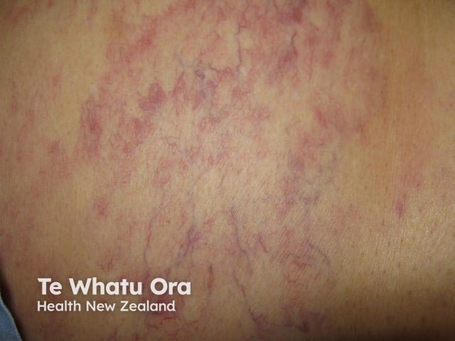
Telangiectatic intravascular lymphoma

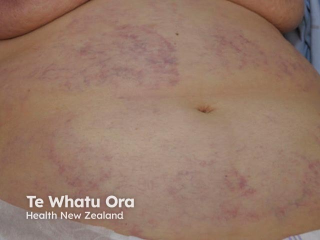
Telangiectatic intravascular lymphoma

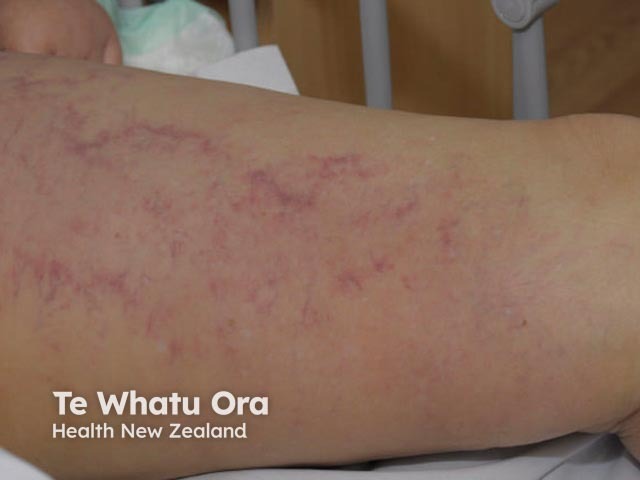
Telangiectatic intravascular lymphoma
Who gets intravascular lymphoma?
Intravascular lymphoma is rare, and its true incidence is unknown. The most typical age of diagnosis is 50–70 years of age. It occurs equally in both men and women [1–3].
The cutaneous variant of intravascular lymphoma is mostly diagnosed in young adult women [1]. There are Western and Asian forms of the disease.
What causes intravascular lymphoma?
Intravascular lymphoma is caused by the proliferation of neoplastic lymphoid cells within capillaries and post-capillary venules. These neoplastic cells are usually mature CD20+ B cells (CD20 is an antigen on the surface of some B cells), but there have been rare cases of T-cell or (NK)-cell intravascular lymphoma [2].
What are the clinical features of intravascular lymphoma?
The presentation of intravascular lymphoma is varied. Typically, and unlike other forms of lymphoma, there is no tumour mass or detectable circulating lymphoma cells.
- Constitutional B symptoms, such as low-grade fever, night sweats, and weight loss, affect the majority of patients.
- Lesions in the cutaneous variant of intravascular lymphoma are similar to cutaneous lesions in the visceral form [4].
- Rapidly progressing neurological signs include dementia, neuropathy, and stroke.
- Cutaneous features that are present in 40% of all patients with intravascular lymphoma include [1,4]:
- Nodules and plaques (49%)
- Red and grey macules
- Lesions on the legs (particularly the anterior inner aspect of the thighs) and trunk.
Other cutaneous features seen in intravascular lymphoma include:
- Dermal oedema (28%) and telangiectasia (20%)
- Painful lesions (24%).
Lymphoma is found in other organs in about 30% of patients with cutaneous features [1]. The spleen, liver, and bone marrow are less frequently involved.
What are the complications of intravascular lymphoma?
Intravascular lymphoma soon results in multi-organ dysfunction, due to bone marrow, renal, hepatosplenic (liver and spleen), and CNS involvement. The disease is commonly fatal within a few months of diagnosis.
How is intravascular lymphoma diagnosed?
Intravascular lymphoma is diagnosed by deep skin biopsy of cutaneous lesions or by random biopsies of apparently healthy skin.
- Ideally, select a cherry angioma to biopsy if present (neoplastic cells can become entrapped in these) [5].
- Reactive perivascular infiltrates in the form of small lymphocytes, and plasma cells accompany abnormal lymphoid cells in the cutaneous capillaries.
- If cutaneous biopsies are non-diagnostic, a biopsy should be carried out on other organs suspected of involvement.
- Immunophenotyping of intravascular lymphoid cells (using CD20 markers and others) is required to differentiate B cell intravascular lymphoma from rarer T-cell or NK-cell lymphoma subtypes.
- Imaging should include magnetic resonance imaging (MRI) for suspected CNS involvement.
- Non-specific blood findings may include [6]:
- Increased lactate dehydrogenase and beta-2 microglobulin (in 80–90% of patients)
- Anaemia (65%)
- Raised erythrocyte rate (ESR) (43%)
- Altered renal, hepatic, or thyroid function (15–20%)
- Monoclonal proteins (14%).
What is the differential diagnosis for intravascular lymphoma?
Several other disorders may be considered in patients who present with the cutaneous features of intravascular lymphoma. These disorders include:
- Thrombophlebitis — this has no telangiectasia or systemic features
- Thrombophlebitis migrans — the affected areas migrate and mainly affect lower legs
- Erysipelas— this causes fever and painful lymphadenopathy (patients with intravascular lymphoma are sub-febrile)
- Erythema nodosum — this is usually found on the anterior shins
- Polyarteritis nodosa — this is characterised by lower leg ulceration and nodules in an arterial distribution; it does not favour the thighs
- Livedo racemosa — this may be accompanied by ulceration and scarring
- Septic emboli — these present as tender inflammatory papules that tend to occur on the digits.
Other inflammatory or immune reactions of the lymph nodes produce activated lymphocytes in the post-capillary venules without any constitutional symptoms, blood derangement, or organ dysfunction.
Intralymphatic histiocytosis is a reactive condition of macrophages, associated with rheumatoid arthritis. It may present with lesions that are clinically similar to intravascular lymphoma and has an intravascular proliferation of large cells. It can be differentiated from intravascular lymphoma on immunophenotyping, which will demonstrate macrophage-specific markers and the absence of lymphoid markers [7].
What is the treatment for intravascular lymphoma?
There are no published guidelines or evidence from controlled trials for the treatment of intravascular lymphoma.
The general expert consensus is that patients with intravascular lymphoma should be considered to have disseminated disease. The majority of patients are treated with intensive chemotherapy; particularly, a combination of the recombinant anti-CD20 drug rituximab, cyclophosphamide, doxorubicin (hydroxydaunomycin), vincristine (Oncovin®) and systemic steroids, such as prednisone or prednisolone (R-CHOP) [1].
Those with a single cutaneous lesion can be considered for treatment with radiotherapy, with or without chemotherapy.
Clinical follow-up is mandatory to detect disease progression and involvement of other organs [4].
What is the outcome for intravascular lymphoma?
Intravascular lymphoma often has a fatal course due to late diagnosis and advanced disease at the time of diagnosis. Biopsy of the skin lesions enables early diagnosis.
In general, patients with the cutaneous variant of intravascular lymphoma have better outcomes than patients who have involvement of other organs (10% of patients with the cutaneous variant have a fatal outcome versus 85% of patients with other organ involvement). The morphology of the skin lesions does not predict the clinical course or prognosis of the lymphoma; however, multiple cutaneous lesions are associated with a worse outcome than single lesions [1].
References
- Ferreri AJ, Campo E, Seymour JF, et al. Intravascular lymphoma: clinical presentation, natural history, management and prognostic factors in a series of 38 cases, with special emphasis on the 'cutaneous variant'. Br J Haematol 2004; 127: 173–83. DOI: 10.1111/j.1365-2141.2004.05177.x. PubMed
- Murase T, Yamaguchi M, Suzuki R, et al. Intravascular large B-cell lymphoma (IVLBCL): a clinicopathologic study of 96 cases with special reference to the immunophenotypic heterogeneity of CD5. Blood. 2007; 15; 109: 478–85. DOI: 10.1182/blood-2006- 01-021253. Journal
- Shimada K, Matsue K, Yamamoto K, et al. Retrospective analysis of intravascular large B-cell lymphoma treated with rituximab-containing chemotherapy as reported by the IVL study group in Japan. J Clin Oncol 2008: 3189–95. DOI: 10.1200/JCO.2007.15.4278. PubMed
- Röglin J, Böer A. Skin manifestations of intravascular lymphoma mimic inflammatory diseases of the skin. Br J Dermatol 2007; 157: 16–25. DOI: 10.1111/j.1365-2133.2007.07954.x. PubMed
- Adachi Y, Kosami K, Mizuta N, et al. Benefits of skin biopsy of senile hemangioma in intravascular large B-cell lymphoma: A case report and review of the literature. Oncol Lett 2014; 7: 2003–6. DOI: 10.3892/ol.2014.2017. PubMed
- Ponzoni M, Ferreri AJ, Campo E, et al. Definition, diagnosis, and management of intravascular large B-cell lymphoma: proposals and perspectives from an international consensus meeting. J Clin Oncol. 2007; 25: 3168. DOI: 10.1200/JCO.2006.08.2313. PubMed
- Requena L, El-Shabrawi-Caelen L, Walsh SN, et al. Intralymphatic histiocytosis. A clinicopathologic study of 16 cases. Am J Dermatopathol 2009; 31:140–51. DOI: 10.1097/DAD.0b013e3181986cc2. PubMed
On DermNet
- Lymphoma
- Vascular skin problems
- Cutaneous B-cell lymphoma
- Telangiectasia
- Vascular proliferations and abnormalities of blood vessels
- Skin manifestations of haematological diseases
Other websites
- Intravascular large call lymphoma — UpToDate
- Intravascular large B-cell lymphoma — Cancer Therapy Advisor
- Non-Hodgkin lymphoma — Medscape
