

Idiopathic guttate hypomelanosis dermoscopy — extra information
Introduction Dermoscopic features Differential diagnoses Histological explanation
What is idiopathic guttate hypomelanosis?
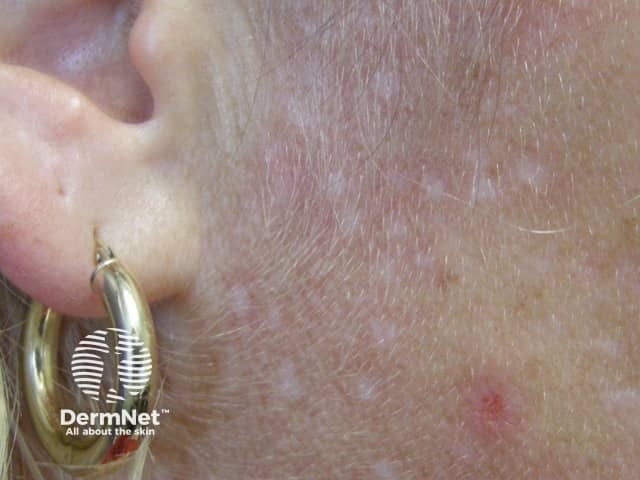
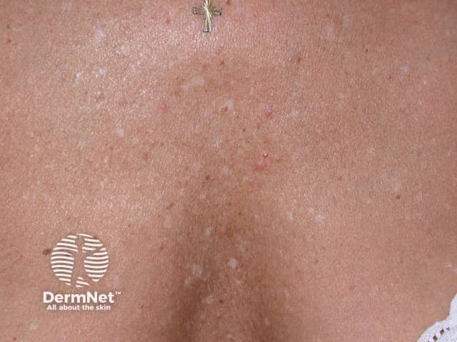
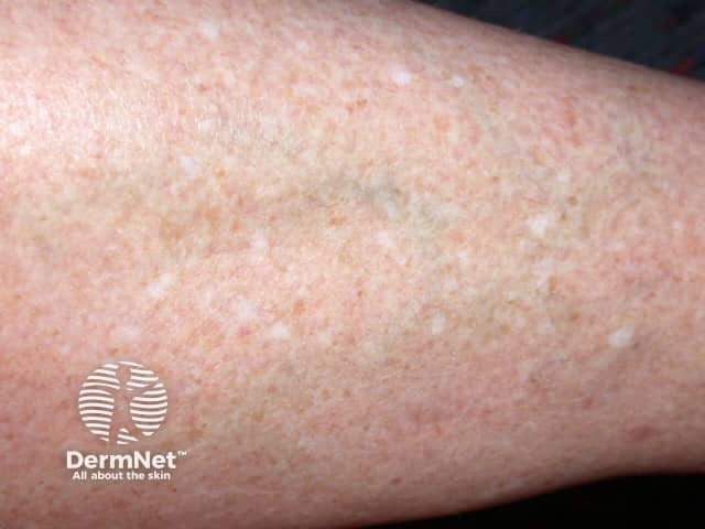
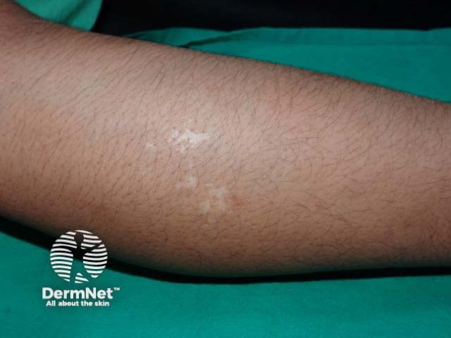
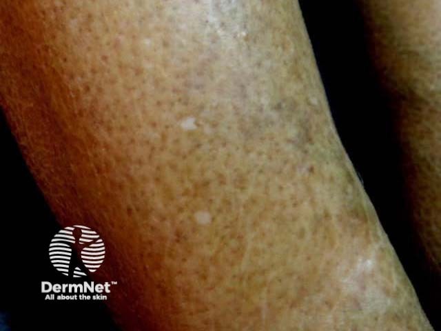
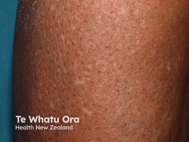
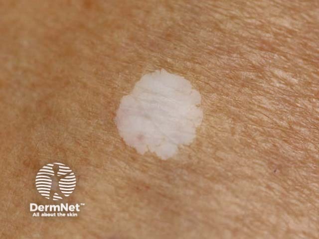
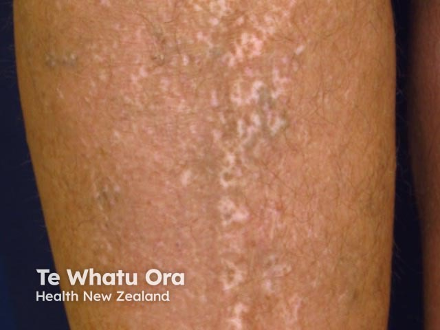
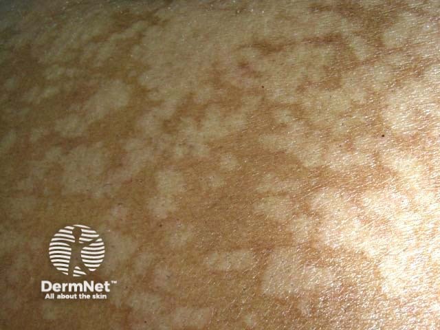
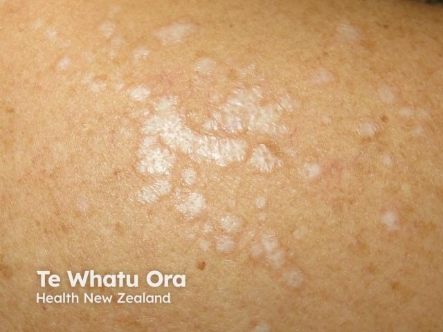
Idiopathic guttate hypomelanosis is a common acquired form of leukoderma characterised by flat porcelain-white macules on sun-exposed areas. The classical sites are the forearms, legs and trunk.
Clinical features of idiopathic guttate hypomelanosis
What are the dermoscopic features of idiopathic guttate hypomelanosis?
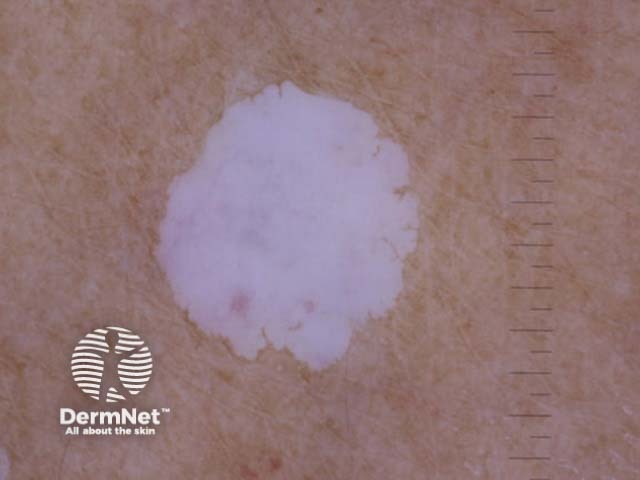
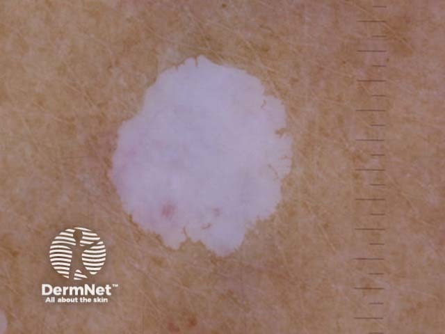
The dermoscopic features of idiopathic guttate hypomelanosis are characterised by white structureless areas in which the pigment network is absent. They extend peripherally with irregular borders and shapes.
White structureless areas appear to ‘glow’ due to total loss of melanocytes in the epidermis. This glow is not as uniform as the glow seen in the dermoscopy of vitiligo and there may be various shades of white.
Metaphoric terms such as amoeboid, feathery, petaloid and nebuloid shapes have been described [3,4].
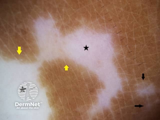
- Amoeboid pattern: white structureless area with a pseudopod-like periphery resembling an amoeba
- Feathery pattern: white structureless area with featherlike linear extensions and irregular margins
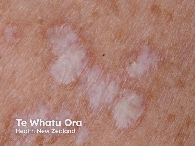
- Petaloid pattern: the white structureless areas resemble the petals of a flower
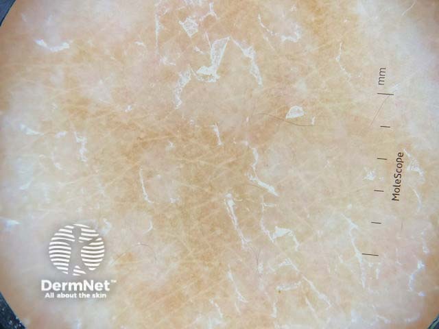
- Nebuloid pattern: the white structureless areas have indistinct borders like a cloud. Other descriptions include ‘cloudy sky-like’ and ‘cloudy’ patterns
- Perifollicular, perilesional or diffuse pigmentation may be observed with occasional red dots [4].
Dermoscopic features of idiopathic guttate hypomelanosis
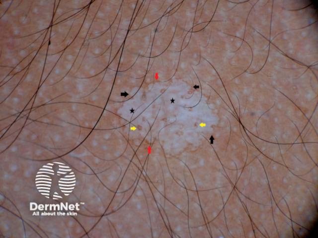
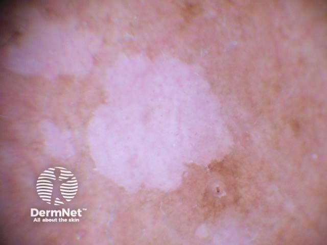
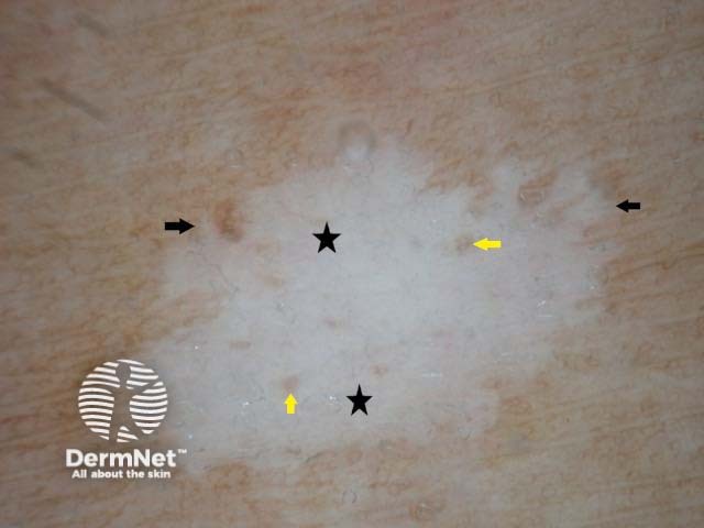
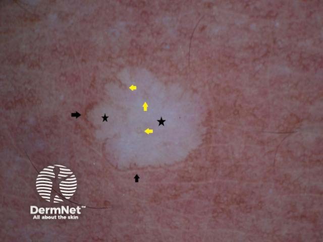
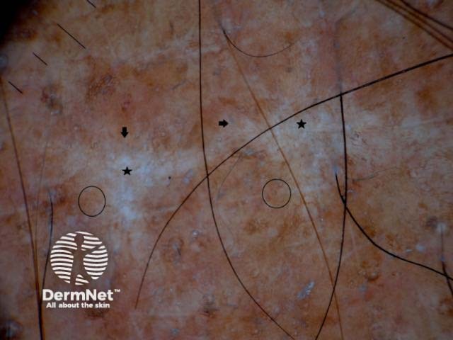
Figure legends Figures 1,2. Amoeboid pattern. Diffuse white structureless area (black stars) with well-defined borders which are extending like pseudopods (black arrows), hence the name. Note the perifollicular (yellow arrows) and perilesional (red arrows) pigmentation.
Figure 3. Feathery pattern. White structureless area (black stars) which extends peripherally in linear strands (black arrows) like a feather. Perifollicular pigmentation (yellow arrows) is well appreciated.
Figure 4. Petaloid pattern with a white structureless area (black stars) and peripheral extensions resembling petals. Perifollicular (yellow arrows) and perilesional (black arrows) pigmentation are also noted.
Figure 5. Nebuloid pattern. White areas (black stars) with indistinct borders (black arrows) and subtle pigmentation (black circles).
Macro and dermoscopy images of idiopathic guttate hypomelanosis
What is the dermoscopic differential diagnosis of idiopathic guttate hypomelanosis?
The dermoscopic differential diagnosis for idiopathic guttate hypomelanosis include guttate vitiligo, pityriasis versicolor and lichen sclerosus.
- Vitiligo dermoscopy: white structureless areas with absent or reduced pigment network, that appear to glow due to total loss of melanocytes in the epidermis. Red dots and linear vessels are not seen See Dermoscopy of vitiligo.
- Pityriasis versicolor dermoscopy: white structureless areas, persisting pigment network, and white scale without a glow. The scale is prominent in the skin lines and separates into lines when the lesion is stretched. See Dermoscopy of pityriasis versicolor
- Lichen sclerosus dermoscopy: comedo-like openings, telangiectasia, perpendicular white lines on polarised dermoscopy, blue-grey peppering and black dots. See Dermoscopy of lichen sclerosus
Dermoscopic differential diagnosis for idiopathic guttate hypomelanosis
What is the histological explanation of idiopathic guttate hypomelanosis?
The histopathology of guttate hypomelanosis shows hyperkeratosis, flattening of rete ridges, epidermal atrophy and acanthosis. There are areas of absent and retained melanocytes; these may explain the white structureless areas and the perifollicular and perilesional pigmentation [5].
References
- Podder I, Sarkar R. Idiopathic guttate hypomelanosis: An overview. Pigment Int [serial online] 2018 [cited 2019 Dec 15];5:83–90. Available from: http://www.pigmentinternational.com/text.asp 2018/5/2/83/247503
- Shin MK, Jeong KH, Oh IH, Choe BK, Lee MH. Clinical features of
idiopathic guttate hypomelanosis in 646 subjects and association with other aspects of photoaging. Int J Dermatol. 2011 Jul; 50(7):798–805. doi:10.1111/j.1365-4632.2010.04743.x. PubMed PMID: 21699514. - Ankad BS, Beergouder SL. Dermoscopic evaluation of idiopathic guttate hypomelanosis: A preliminary observation . Indian Dermatol Online J [serial online] 2015 [cited 2019 Dec 15];6:164–7. Available from: http://www.idoj.in/text.asp?2015/6/3/164/156383
- Errichetti E, Stinco G. Dermoscopy of idiopathic guttate hypomelanosis. J Dermatol. 2015 Nov;42(11):1118–9. doi: 10.1111/1346-8138.13035. Epub 2015 Jul 27. PubMed PMID:26212033
- Joshi R. Skip areas of retained melanin: a clue to the histopathological diagnosis of idiopathic guttate hypomelanosis. Indian J Dermatol. 2014 Nov; 59(6):571–4. doi: 10.4103/0019-5154.143516. PubMed PMID: 25484386; PubMed Central PMCID: PMC4248493.
On DermNet
- Idiopathic guttate hypomelanosis
- Pigmentation disorders
- Dermatological conditions in skin of colour
- Skin pigmentation problems