Main menu
Common skin conditions

NEWS
Join DermNet PRO
Read more
Quick links
Hypergranulotic dyscornification — extra information
Hypergranulotic dyscornification
DermNet September 2021. Copy edited by Gus Mitchell.
Introduction Demographics Causes Clinical features Diagnosis Differential diagnoses Treatment
What is hypergranulotic dyscornification?
Hypergranulotic dyscornification is a reaction pattern seen on histology following excision of a benign keratotic skin lesion.
Who gets hypergranulotic dyscornification?
Hypergranulotic dyscornification has been rarely reported. In the one large pathology series, there was a female predominance of almost 2:1. All reported cases have been adults with a mean age at diagnosis of 57 years. There is no information regarding racial distribution at this time.
What causes hypergranulotic dyscornification?
The cause of hypergranulotic dyscornification is unknown. Theories include a possible keratin mutation or other disorder of keratinisation and epidermal maturation.
What are the clinical features of lesions associated with hypergranulotic dyscornification?
Hypergranulotic dyscornification has almost exclusively been seen in lesions described as:
- Solitary scaly
- Waxy tan colour
Most have been excised from the lower limbs, followed by the trunk. Upper limbs, head, and neck are less commonly reported sites.
How is hypergranulotic dyscornification diagnosed?
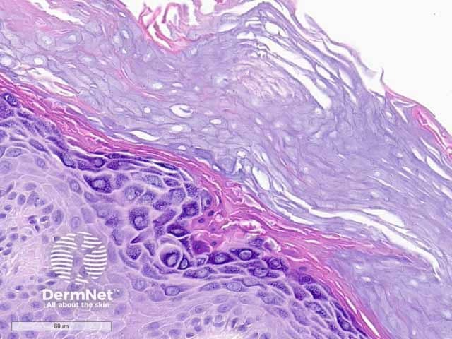
Hypergranulotic dyscornification is diagnosed on histological examination of an excised skin lesion [see Hypergranulotic dyscornification pathology].

Hypergranulotic dyscornification: histology high power
What is the clinical differential diagnosis for hypergranulotic dyscornification?
- Seborrhoeic keratosis
- Non-melanoma skin cancer or actinic keratosis
- Viral wart
What is the treatment for hypergranulotic dyscornification?
As hypergranulotic dyscornification is only diagnosed after excisional biopsy, further treatment is not required. All reported lesions showing hypergranulotic dyscornification have been benign to date.
Bibliography
- Gesheva A, Pitch M, Rosamilia L, Hossler E. Keratotic papule on the abdomen. Cutis. 2020;105(5):222–31. Journal
- Roy SF, Ko CJ, Moeckel GW, Mcniff JM. Hypergranulotic dyscornification: 30 cases of a striking epithelial reaction pattern. J Cutan Pathol. 2019;46(10):742–7. doi:10.1111/cup.13522. PubMed
On DermNet
