Main menu
Common skin conditions

NEWS
Join DermNet PRO
Read more
Quick links
Granulomatous dermatitis — extra information
Granulomatous dermatitis
Author: Dr Susan Simpkin, Dermatology Registrar, Green Lane Clinical Centre, Auckland, New Zealand, 2012.
Introduction
Clinical features
Diagnosis - interstitial granulomatous dermatitis
Introduction - Palisading neutrophilic granulomatous dermatitis
Diagnosis - palisading neutrophilic granulomatous dermatitis
Clinical associations
Treatment
Interstitial granulomatous drug reaction
What is granulomatous dermatitis?
Granulomatous dermatitis describes several disorders characterised by their histological appearance.
- Interstitial granulomatous dermatitis (IGD)
- Palisading neutrophilic granulomatous dermatitis (PNGD)
- Interstitial granulomatous drug reaction (IGDR)
Interstitial granulomatous dermatitis
Interstitial granulomatous dermatitis is a rare skin disorder in which there is a particular pattern of granulomatous inflammation.
The classic original clinical description of interstitial granulomatous dermatitis was of linear erythematous palpable cords on the lateral aspects of the trunk, called 'the rope sign'. However, several different types of rash have since been described with the same histological appearance.
What are the clinical features of interstitial granulomatous dermatitis?
The features of interstitial granulomatous dermatitis are variable.
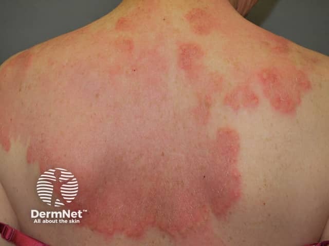
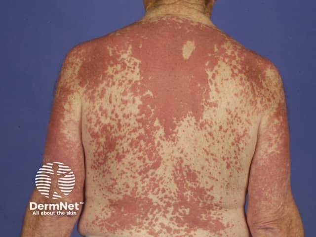
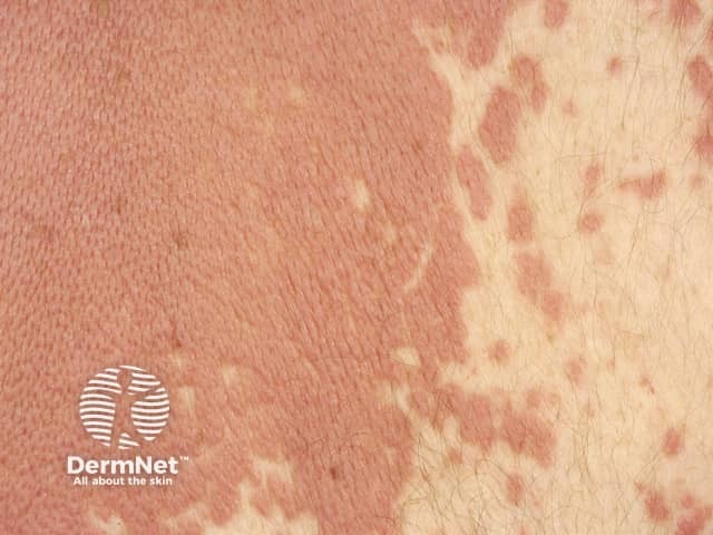
- Red or skin-coloured patches, papules and plaques
- The shape of the lesions may be round, annular or cord-like.
- Lesions wax and wane, and may vary in size and shape over days to months.
- They are usually symptomless, but some patients complain of mild itch or burning sensation.
- The lesions tend to be symmetrically distributed on the trunk, but proximal limbs may also be affected.
- It most commonly affects middle-aged women.
- Many affected patients also suffer from autoimmune disease.

Interstitial granulomatous dermatitis

Interstitial granulomatous dermatitis

Interstitial granulomatous dermatitis
How is interstitial granulomatous dermatitis diagnosed?
Interstitial granulomatous dermatitis is diagnosed by a pathologist on examining a skin biopsy. The characteristic histological features of interstitial granulomatous dermatitis are:
- Dense histiocytic inflammation in the reticular (lower) dermis
- Sparse neutrophils and eosinophils
- Perivascular and interstitial lymphocytes
- Interstitial histiocytes (between the collagen bundles) or palisaded histiocytes (lined up perpendicular to fragmented central collagen).
- Focal degeneration of collagen, which may be surrounded by space (floating sign)
- Few giant cells.
How does interstitial granulomatous dermatitis compare to granuloma annulare?
Granuloma annulare and interstitial granulomatous dermatitis may appear similar clinically and histologically. Granuloma annulare also presents with papules and plaques, but these are typically on the back of the hands or feet, whereas the trunk is a more common site for interstitial granulomatous dermatitis. Granuloma annulare is less often associated with autoimmune diseases.
The characteristic histological features of granuloma annulare are:
- Histiocytes located in the upper dermis
- Rare or absent neutrophils and eosinophils
- Abundant mucin [1].
Palisading neutrophilic granulomatous dermatitis
Palisading neutrophilic granulomatous dermatitis was first described as crusted umbilicated papules on the elbows arising in patients with rheumatoid arthritis (Winkelmann granuloma) and eosinophilic granulomatosis with polyangiitis (Churg-Strauss syndrome).
Several different types of rash have been described with the same histological appearance including annular plaques on the trunk. Lesions are often tender and may ulcerate.
How is palisading neutrophilic granulomatous dermatitis diagnosed?
Palisading neutrophilic granulomatous dermatitis is diagnosed on skin biopsy. The characteristic histological features of palisading neutrophilic granulomatous dermatitis are:
- Intense neutrophilic infiltrate and nuclear dust
- Interstitial histiocytic infiltrate
- Collagen degeneration
- Leukocytoclastic vasculitis
- Variable histological appearance depending on the stage of the eruption.
Palisading granulomas seen have also been described as miniature ‘Churg-Strauss granulomas’ or flame figures, with degenerated collagen enveloped by eosinophils resembling a flame. Flame figures are also seen in the pathology of Wells syndrome.
What are the clinical associations of granulomatous dermatitis?
Several conditions have been noted to arise in association with interstitial granulomatous dermatitis. There is less information about associations with palisading neutrophilic granulomatous dermatitis.
Autoimmune diseases and conditions
Both forms of granulomatous dermatitis have often arisen in people with other conditions considered autoimmune in origin, implying an immune-complex mechanism may be involved in the pathogenesis [2]. These have included the following conditions.
- Rheumatoid and non-rheumatoid arthritis, characteristically symmetrical and involving the fingers, wrists, elbows, and shoulders, is the most common association. Arthritis (inflamed joints) or arthralgias (painful joints) may occur before, during or years after the onset of the skin lesions in about 50% of published cases of interstitial granulomatous dermatitis [3].
- The systemic lupus erythematosus (SLE)
- Primary antiphospholipid syndrome
- Churg-Strauss syndrome
- Thyroiditis
- Vitiligo
In a literature review of 15 patients with interstitial granulomatous dermatitis, autoantibodies identified on blood testing included rheumatoid factor (RhF), antinuclear factor (ANA), thyroglobulin, SS-A Histone, DNA histone and ANCA [4].
Malignancy
There are some reports of association of malignancy with interstitial granulomatous dermatitis. In one case, the lesions cleared after lung cancer was treated [5]. Rare associations of granulomatous dermatitis with leukaemia [6], lymphoma, breast cancer, hypopharyngeal squamous cell carcinoma and endometrial neoplasia have been reported.
How is granulomatous dermatitis treated?
Granulomatous dermatitis typically flares and remits. Successful treatments have included:
One case associated with SLE responded well to systemic steroids after 15 days [2].
Although tumour necrosis factor (TNF)-alpha inhibitors have been recently described as inducing interstitial granulomatous dermatitis [7], etanercept has been used in interstitial granulomatous dermatitis associated with rheumatoid arthritis with complete skin clearance and improvement in arthritis [8].
Interstitial granulomatous drug reaction
Interstitial granulomatous dermatitis induced by medications is known as an interstitial granulomatous drug reaction. It is thought to be a distinct clinical and pathological entity. It presents as annular plaques, and nodules on the trunk, arms, medial thighs and skin folds. The rash resolves when the responsible drug is withdrawn.
How is an interstitial granulomatous drug reaction diagnosed?
Interstitial granulomatous drug reaction is diagnosed by skin biopsy. The characteristic histological features of interstitial granulomatous drug reaction are:
- Basal epidermal cell vacuolisation
- Lichenoid changes with eosinophils
- Absence of neutrophils
- In some cases, palisaded granulomatous changes associated with collagen necrosis and floating sign.
Medications that have been reported to be implicated in interstitial granulomatous drug eruption include:
-
TNF-alpha inhibitors (infliximab, adalimumab, etanercept) [7]
- Beta-blockers
- Calcium channel blockers
- Angiotensin-converting enzyme inhibitors
- Lipid-lowering agents
- Statins (HMG-CoA reductase inhibitors)
- Frusemide
- Antidepressants
- Anticonvulsants [4].
References
- García-Rabasco A, Esteve-Martínez A, Zaragoza-Ninet V, Sánchez-Carazo JL, Alegre-de-Miquel V. Interstitial Granulomatous Dermatitis in a Patient With Lupus Erythematosus. Am J Dermatopathol 2011; 33(8):871–2. Journal
- Blaise S, Salameire D, Carpentier PH. Interstitial granulomatous dermatitis: a misdiagnosed cutaneous form of systemic lupus erythematosus? Clin Exp Dermatol 2008; 33: 712–4. PubMed
- Peroni A, Colato C, Schena D, Gisondi P, Girolomoni G. Interstitial granulomatous dermatitis: a distinct clinical entity with characteristic histological and clinical pattern. Br J Dermatol. 2012; 66(4):775–83. PubMed
- Verneuil L, Dompmartin A, Comoz F, Pasquier CJ, Leroy D. Interstitial granulomatous dermatitis with cutaneous cords and arthritis: A disorder associated with autoantibodies. J Am Acad Dermatol 2001; 45: 286-91. PubMed
- Schreckenberg C, Asch PH, Sibilia J, Walter S, Lipsker D, Heid E, et al. Dermatite granulomateuse interstitielle et polyarthrite rhumatoïde paranéoplasiques révélatrices d’un cancer du poumon. Ann Dermatol Venereol 1998; 125: 585-8. PubMed
- Swing DC, Sheehan DJ, Sangüeza OP, Woodruff RW. Interstitial granulomatous dermatitis secondary to acute promyelocytic leukemia. Am J Dermatopathol 2008; 30 (2): 197–9. PubMed
- Deng A, Harvey V, Sina B, Strobel D, Badros A et al. Interstitial granulomatous dermatitis associated with the use of tumor necrosis factor alpha inhibitors. Arch Dermatol. 2006; 142: 198–202. PubMed
- Zoli A, Massi G, Pinnelli M, Cuccio C, Castri F et al. Interstitial granulomatous dermatitis in rheumatoid arthritis responsive to etanercept. Clin Rheumatol (2010); 29: 99–101. PubMed
On DermNet
- Granuloma annulare
- Urticaria and urticaria-like conditions
- Skin signs of rheumatic disease
- Interstitial granulomatous dermatitis pathology
Other websites
- Non-Infectious Granulomatous Diseases of the Skin: 5. Are Interstitial Granulomatous Dermatitis and Palisaded Neutrophilic Granulomatous Dermatitis Distinct Conditions with Overlapping Features and Triggers, or Part of a Single Disease Spectrum? – Medscape Dermatology News
