Main menu
Common skin conditions

NEWS
Join DermNet PRO
Read more
Quick links
Eccrine carcinoma pathology — extra information
Lesions (cancerous) Diagnosis and testing
Eccrine carcinoma pathology
Author: Assoc Prof Patrick Emanuel, Dermatopathologist, Auckland, New Zealand, 2013.
Histology of eccrine carcinoma
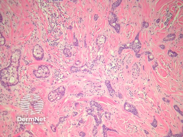
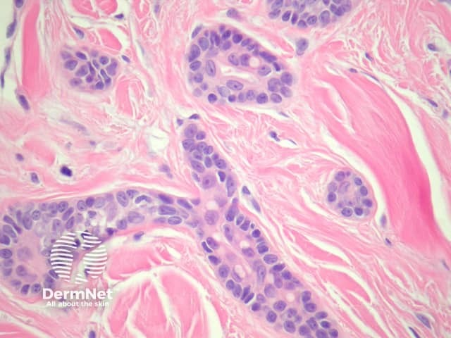
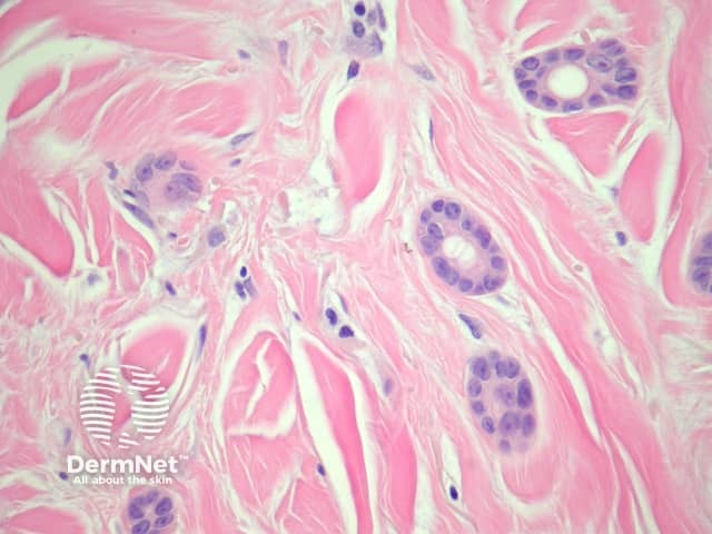
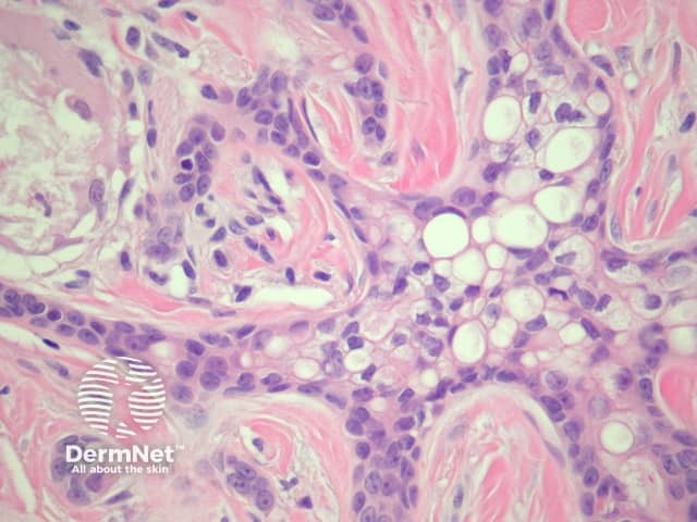
Sections of eccrine carcinoma show a tumour composed of basaloid cells infiltrating the dermis (figure 1). The tumour forms tubules and glands (figures 2-4) which infiltrate a sclerotic dermis. Eccrine differentiation is readily seen at high power (best seen in figures 2 and 3). Clear cell change may be seen (figure 4).

Figure 1

Figure 2

Figure 3

Figure 4
Special studies for eccrine carcinoma
Immunohistochemical studies can be used to demonstrate glandular differentiation (CEA) in eccrine carcinoma, but are not needed if the morphology is classic. Some cases resemble breast cancer. Focal p63 and CK5/6 positivity is thought to favour a primary eccrine carcinoma.
Differential diagnosis of eccrine carcinoma
Metastatic breast cancer – Clinical correlation may be needed to distinguish primary eccrine carcinoma from metastatic breast carcinoma. Immunohistochemical studies (see above) can be helpful
Microcystic adnexal carcinoma (MAC) – Some authors consider eccrine carcinoma to be a form of MAC. MAC generally demonstrates some squamous differentiation and keratinizing cystic changes superficially.
.
References
- Pathology of the Skin (Fourth edition, 2012). McKee PH, J. Calonje JE, Granter SR
On DermNet
