Main menu
Common skin conditions

NEWS
Join DermNet PRO
Read more
Quick links
Dermoscopy of leprosy — extra information
Infections Diagnosis and testing
Dermoscopy of leprosy
Author: Professor Balachandra Ankad, Department of Dermatology, S Nijalingappa Medical College, Bagalkot-587103, Karnataka, India. Copy edited by Gus Mitchell. January 2021.
Introduction
Clinical features
Dermoscopic features
Differential diagnoses
Histological explanation
What is leprosy?
Leprosy is a chronic granulomatous disorder of the skin and nerves usually associated with Mycobacterium leprae infection.
What are the clinical features of leprosy?
Leprosy presents as a spectrum of disease ranging from multibacillary to paucibacillary forms. The cardinal clinical features of leprosy are hypopigmented anaesthetic patches on the skin and thickened, tender peripheral nerves.
What are the dermoscopic features of leprosy?
Dermoscopic patterns in leprosy vary with the skin site/location, part of the lesion examined, and the type of lesion eg, flat versus raised.
General dermoscopy features of leprosy
- Focal white areas
- Distorted pigment network
- Reduced density of white dots indicating eccrine and follicular openings
- Hair changes — circle hairs, short or broken hairs, V-like hairs, decreased hair density
- Widened skin cleavage lines
- Occasional scale only seen in infiltrated lesions
- Vascular changes — short, linear, dotted, and branching blood vessels in granulomatous and facial lesions; not seen in extra-facial flat hypopigmented lesions
- Yellow and/or yellow-brown globules — in granulomatous facial lesions and infiltrated extra-facial lesions.
Specific dermoscopy features of leprosy
Tuberculoid leprosy
Yellow-orange and brown-orange globules with linear and branching blood vessels which may be poorly focused. Vessels are very difficult to appreciate in flat hypopigmented extra-facial lesions.
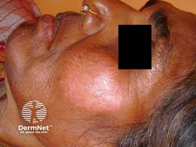
Well-defined erythematous plaque on the cheek in skin of colour
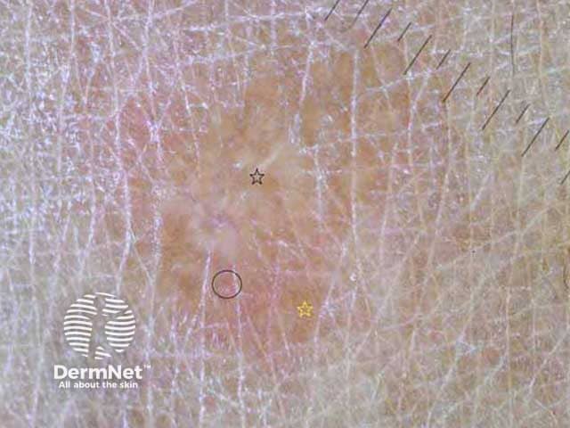
Borderline tuberculoid leprosy
Focal white areas and distorted pigment network with white structureless areas. Yellow-orange and/or brown-yellow globular structures with linear vessels are seen in facial lesions and raised extra-facial lesions.
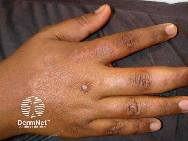
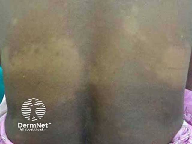
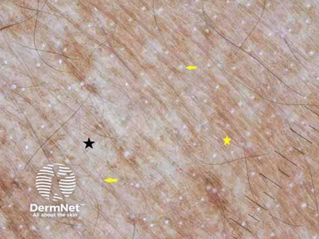
Borderline-borderline leprosy
Focal white areas, distorted pigment network, ill-defined focal red areas with decreased white dots of eccrine and follicular openings. It is difficult to appreciate brown-yellow globules in extra-facial sites in dark skin. A pinkish background can be observed.
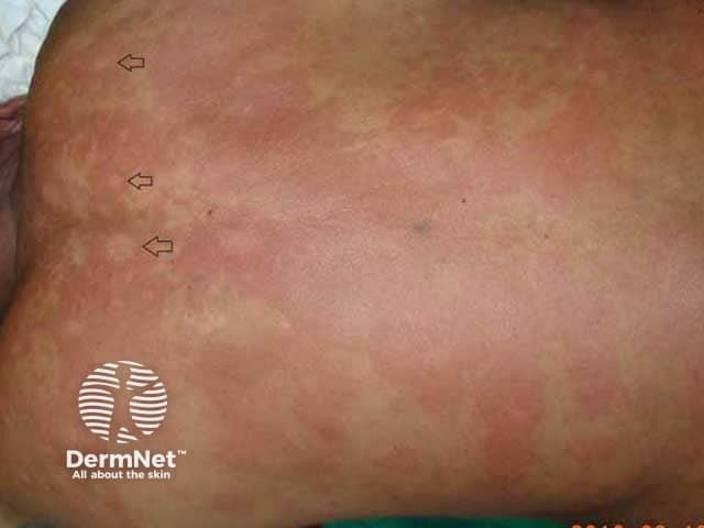
Red lesions of variable size. Characteristic 'Swiss cheese' appearance (black arrows)
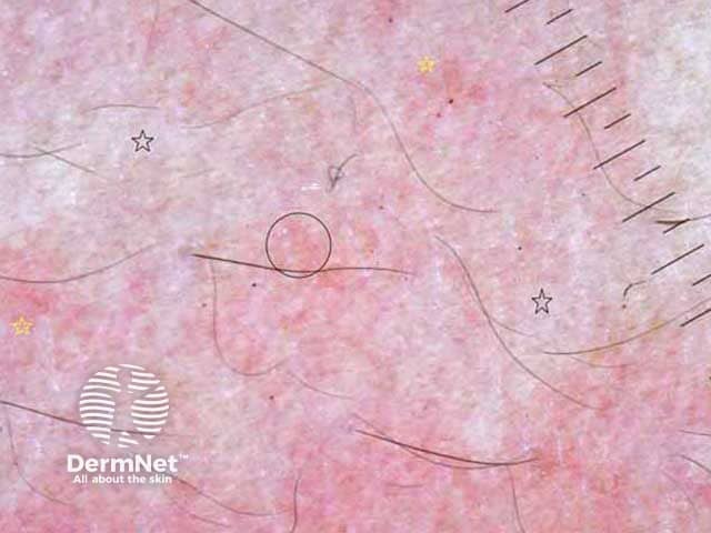
Borderline lepromatous leprosy
Distorted pigment network with reduction in white dots of eccrine and follicular openings. Focal white areas with white chrysalis strands are usually seen. Brown-yellow globules are not easily appreciated. White rosettes have been described in facial lesions and are non-specific, being seen in many other conditions.
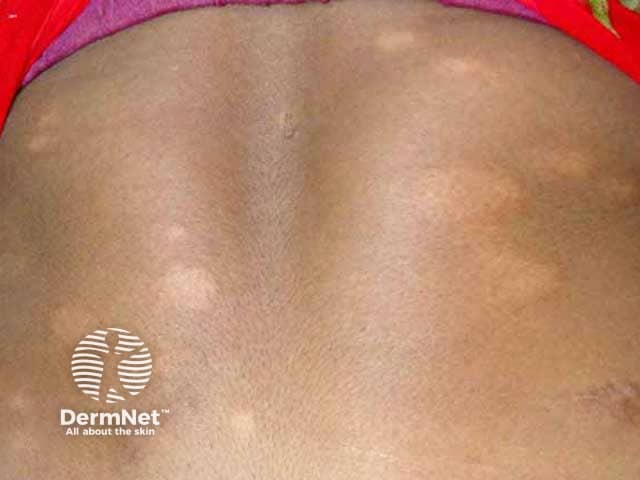
Asymmetrical distribution of erythematous infiltrated plaques in skin of colour
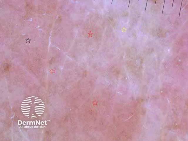
Lepromatous leprosy
Distorted pigment network with chrysalis strands and prominent linear and branching vessels are seen in nodular lesions. Vascular elements are not obvious in patches. Globules are difficult to find.
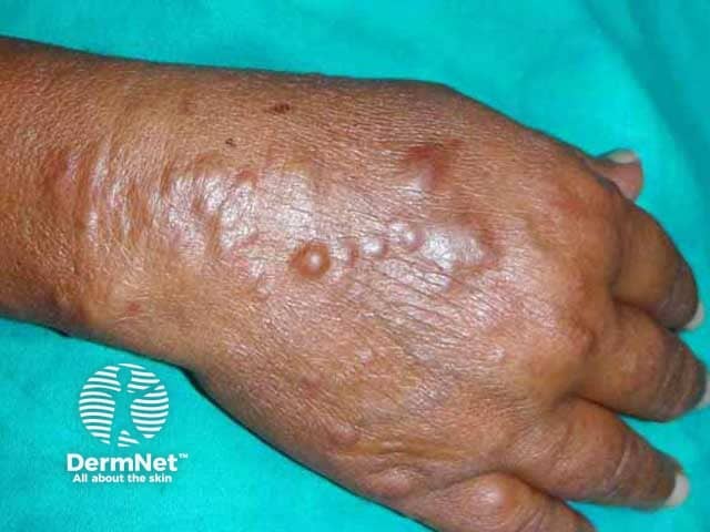
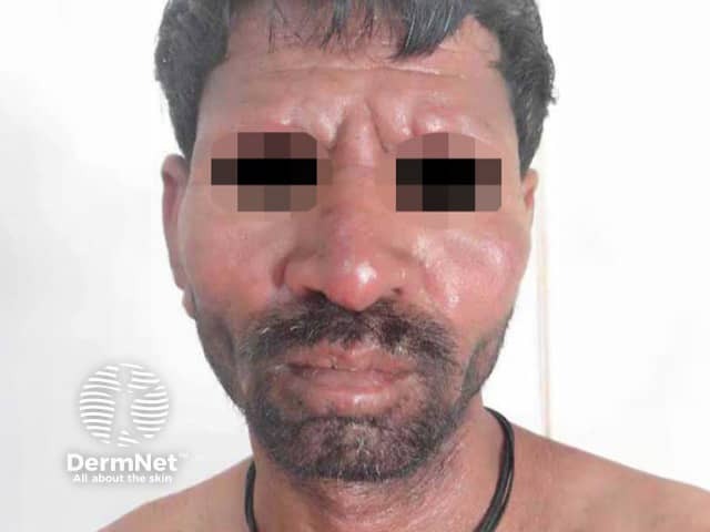
Leonine facies with loss of eyebrows and infiltrated facial skin
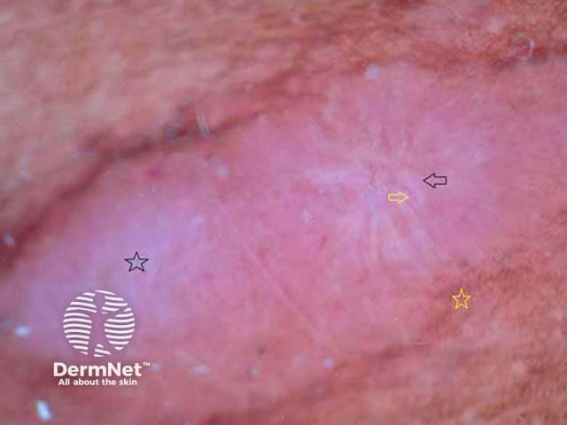
Histoid leprosy
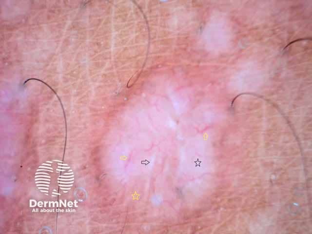
White structureless areas with linear and branching vessels running towards the centre of the lesion, so-called crown vessels. White chrysalis strands and peripheral brownish pigment are typically seen.
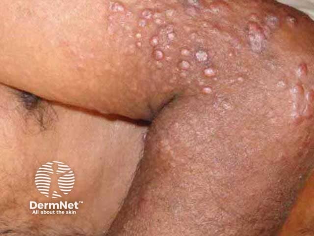
Dome-shaped papules and nodules in skin of colour
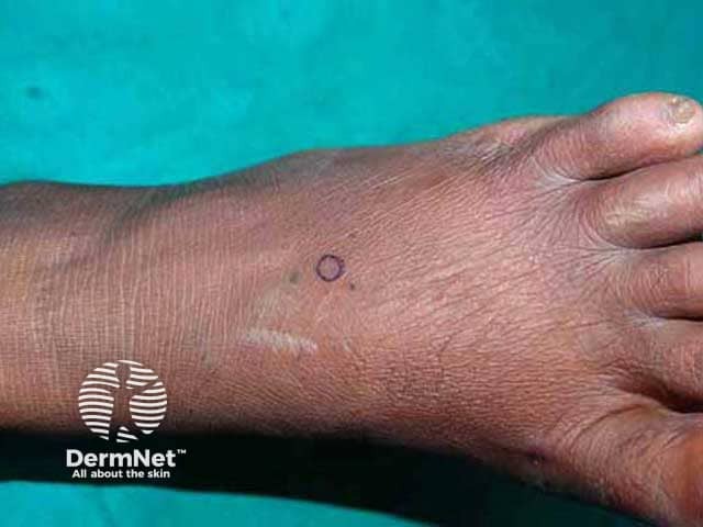
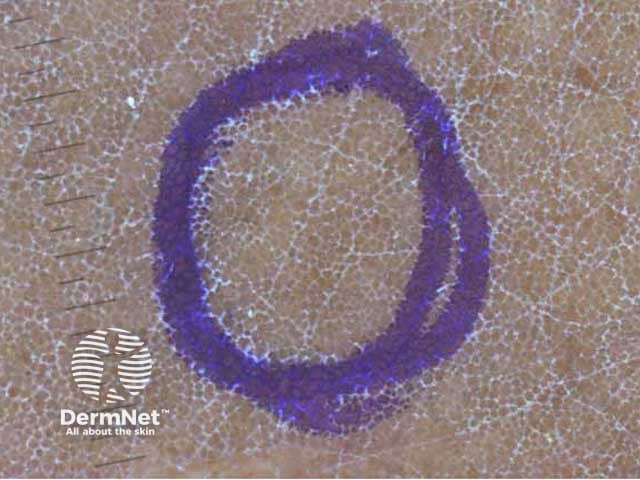
Pure neural leprosy
Prominent skin lines with white scale and changes in the pigment network.
What is the dermoscopic differential diagnosis for leprosy?
- Pityriasis versicolor dermoscopy: diffuse white structureless areas with faint pigment network and fine white scale. No changes seen in hair and vascular elements.
- Hypopigmented mycosis fungoides dermoscopy: short linear spermatozoan-like vessels, focal white scales, brown-yellow structures, and a whitish background.
- Lupus vulgaris dermoscopy: brown-yellow globules, linear and branching vessels, and white chrysalis strands. Follicular plugs and prominent white scale are conspicuous in lupus vulgaris but not in leprosy.
- Sarcoidosis dermoscopy: well-focused linear vessels with orange-yellow globules, scale, and follicular plugs.
What is the histological explanation of the dermoscopic features of leprosy?
Focal white areas and broken pigment networks correspond with decreased basal layer melanin and flattening of rete ridges respectively. The decrease in white dots is due to a reduced number of follicular and eccrine structures. Granulomas are seen particularly on skin biopsy of the tuberculoid forms of leprosy, and are responsible for the brown-yellow globules. Linear and branching vessels are dilated capillaries. White chrysalis strands correlate with dermal collagen [see Leprosy pathology].
Bibliography
- Abadías-Granado I, Navarro-Bielsa A, Gómez-Mateo MC, Bermúdez-Cameo R, Gilaberte Y. Crown vessels and shiny white structures in dermoscopy of histoid leprosy. JAAD Case Rep. 2020;6(11):1147–9. doi:10.1016/j.jdcr.2020.08.040. PubMed Central
- Acharya P, Mathur MC. Clinicodermoscopic study of histoid leprosy: a case series. Int J Dermatol. 2020;59(3):365–8. doi:10.1111/ijd.14731. PubMed
- Ankad BS, Koti VR. Dermoscopic approach to hypopigmentary or depigmentary lesions in skin of color. Clin Dermatol Rev. 2020;4:79–83. doi: 10.4103/CDR.CDR_68_20. Journal
- Ankad BS, Sakhare PS. Dermoscopy of histoid leprosy: a case report. Dermatol Pract Concept. 2017;7(2):63–5. doi:10.5826/dpc.0702a14. PubMed Central
- Ankad BS, Sakhare PS. Dermoscopy of borderline tuberculoid leprosy. Int J Dermatol. 2018;57(1):74–6. doi:10.1111/ijd.13731. PubMed
- Chatterjee M, Neema S. Dermoscopy of pigmentary disorders in brown skin. Dermatol Clin. 2018;36(4):473–85. doi:10.1016/j.det.2018.05.014. PubMed
- Chopra A, Mitra D, Agarwal R, Saraswat N, Talukdar K, Solanki A. Correlation of dermoscopic and histopathologic patterns in leprosy - a pilot study. Indian Dermatol Online J. 2019;10(6):663–8. doi:10.4103/idoj.IDOJ_297_18. PubMed Central
- Errichetti E, Stinco G. Dermatoscopy of granulomatous disorders. Dermatol Clin. 2018;36(4):369–75. doi:10.1016/j.det.2018.05.004. PubMed
- Martin IG, Ankad BS, Errichetti E, Lallas A, Ionnides D, Zaballos P. Bacterial and parasitic infections. In: Lallas A, Errichetti E, Ionnides D (eds). Dermoscopy in General Dermatology. CRC Press, 2018: 188–209.
- Mathur M, Acharya P, Karki A. Visual dermatology: crown vessels in dermoscopy of histoid leprosy. J Cutan Med Surg. 2019;23(3):333. doi:10.1177/1203475419825759. PubMed
- Vinay K, Kamat D, Chatterjee D, Narang T, Dogra S. Dermatoscopy in leprosy and its correlation with clinical spectrum and histopathology: a prospective observational study. J Eur Acad Dermatol Venereol. 2019;33(10):1947–51. doi:10.1111/jdv.15635. PubMed
On DermNet
Other websites
- World Health Organisation (WHO)
- Hansen's Disease (Leprosy) — Centers for Disease Control and Prevention (CDC)
- Lepra (The British Leprosy Relief Association)
- Leprosy — Medscape reference
