

Chondrodermatitis nodularis pathology — extra information
Histology of chondrodermatitis nodularis
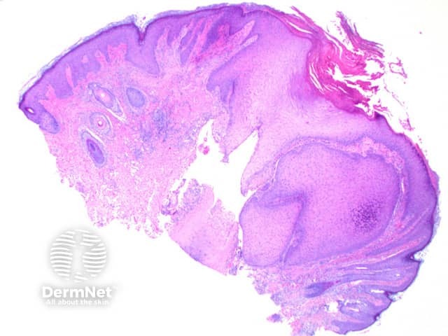
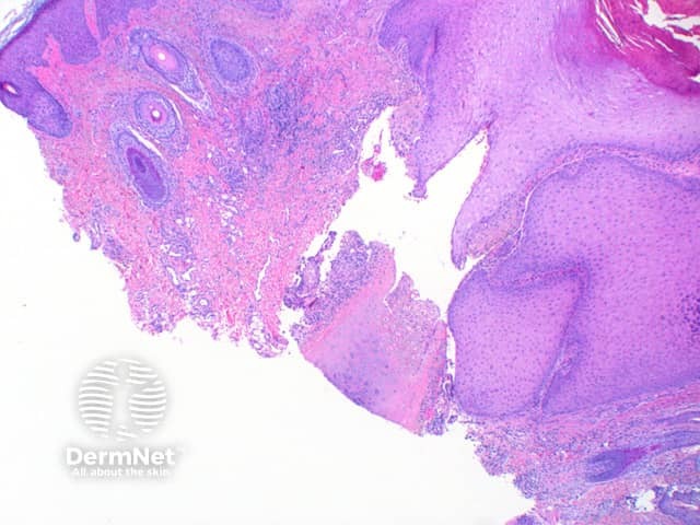
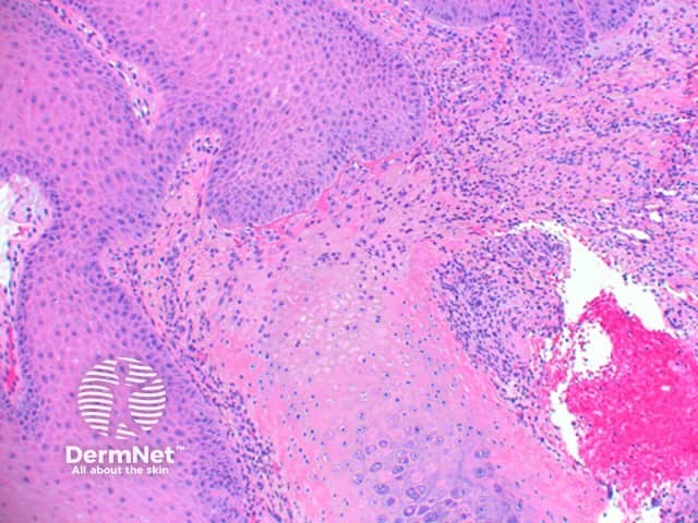
Scanning power view of chondrodermatitis nodularis helicis shows a wedge-shaped dermal alteration extending through to the superficial cartilage (Figure 1). The epidermis may show a focal zone of orthokeratosis and scale crust overlying moderate epidermal hyperplasia (Figure 2). Frequently there will be superficial epidermal ulceration. There is a zone of eosinophilic fibrinoid material overlying the affected area of cartilage, with the blurring of the superficial cartilage and altered staining (Figure 3). There may be adjacent perichondrial thickening.
Differential diagnosis of chondrodermatitis nodularis
Pseudocyst of the auricle: while there may also be hyalinised degeneration above the affected collagen, a non-epithelial lined cystic space forms within the collagen in pseudocyst of the auricle.
References
- Skin Pathology (3rd edition, 2002). Weedon D
- Pathology of the Skin (3rd edition, 2005). McKee PH, J. Calonje JE, Granter SR
On DermNet