Main menu
Common skin conditions

NEWS
Join DermNet PRO
Read more
Quick links
Autonomic denervation dermatitis — extra information
Autonomic denervation dermatitis
Last reviewed: September 2023
Author(s): Dr Amy Cunliffe, Speciality Dermatology Registrar; and Dr Rabi Nambi, Consultant Dermatologist, University Hospitals of Derby and Burton, United Kingdom (2023)
Reviewing dermatologist: Dr Ian Coulson
Edited by the DermNet content department
Introduction
Demographics
Causes
Clinical features
Variation in skin types
Complications
Diagnosis
Differential diagnoses
Treatment
Prevention
Outcome
What is autonomic denervation dermatitis?
Autonomic denervation dermatitis (ADD) is an uncommon localised eczematous eruption around a surgical incision site. It is often associated with cutaneous anaesthesia or hypoesthesia, and may result from iatrogenic nerve damage and autonomic disruption from surgery.
Various nomenclatures, such as traumatic eczematous dermatitis and neuropathy dermatitis, have been used synonymously to describe different entities. Surgery of the Knee, Injury to the infrapatellar branch of the saphenous Nerve, Traumatic Eczematous Dermatitis (SKINTED) refers to a subset of patients who develop autonomic denervation dermatitis following knee surgery.

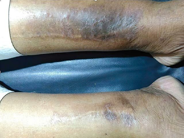
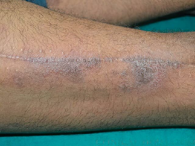
Hyperpigmented eczematous reaction at the site of an orthopaedic operation incision

Hyperpigmented eczematous reaction at the site of an orthopaedic operation incision

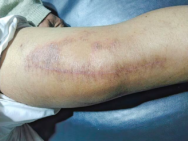
Autonomic denervation dermatitis at the site of a total knee joint replacement skin incision
Who gets autonomic denervation dermatitis?
Data on autonomic denervation dermatitis is limited. At least 82 cases have been reported in the literature but this is likely to be an underestimate of the incidence. It affects both female and male adults, the majority over the age of 50 years.
Autonomic denervation dermatitis most frequently occurs following total knee replacement (TKR) surgery. It has also been reported following other surgical procedures including saphenous vein graft harvesting for coronary artery bypass surgery, knee arthroscopies, fracture fixations of the leg, and amputation.
A small prospective study of 203 consecutive TKR patients reported a 4.4% incidence of autonomic denervation dermatitis.
What causes autonomic denervation dermatitis?
Autonomic denervation dermatitis may result from a denervation injury due to the transection of dermal nerve fibres during surgery and autonomic disruption. The infrapatellar branch of the saphenous nerve is particularly vulnerable during TKR.
Various contributing mechanisms have been speculated such as:
- An altered epidermal barrier function
- Release of neuropeptides during nerve regeneration
- Altered behaviour of keratinocytes
- Local immune dysregulation due to lymphatic trauma resulting in an inflammatory cascade.
What are the clinical features of autonomic denervation dermatitis?
Onset may develop 3 weeks to 3 years following surgery.
Clinical features include:
- A xerotic, scaly erythematous rash, with possible plaques, macules, papules, crusting, and occasionally vesicles
- The rash is localised exclusively to the surgical incision site and the surrounding skin
- Hypoaesthesia or anaesthesia, which may resolve before the onset of the rash
- Pruritus may be present.
Autonomic denervation dermatitis following TKR performed through an anterior midline incision is frequently associated with cutaneous sensory loss in the distribution of the infrapatellar branch of the saphenous nerve. The eczematous eruption is often localised lateral to the midline incision, although it can cross to both sides.
Patients who develop autonomic denervation dermatitis following bilateral TKRs often present with unilateral disease, although bilateral presentations have been reported.
How do clinical features vary in differing types of skin?
There are no reports in the literature of different clinical presentations in various skin types, however erythema is often less apparent in darker skin types, which may affect the appearance of the rash.
What are the complications of autonomic denervation dermatitis?
Autonomic denervation dermatitis may result in:
- Reduction in patient satisfaction following surgery, especially if significant pruritus occurs or rehabilitation is affected
- Secondary infection.
No functional limitations have been reported.
How is autonomic denervation dermatitis diagnosed?
Autonomic denervation dermatitis is diagnosed clinically. Bacterial swabs can be considered to exclude infection.
Skin biopsy is not routinely required for diagnosis. Histology shows subacute or chronic spongiotic dermatitis with a lymphocytic infiltrate.
Patch testing is often negative and is not routinely required. However, a minority of patients test positive to chromate, nickel, or cobalt; the significance of this is unclear.
What is the differential diagnosis for autonomic denervation dermatitis?
- Allergic contact dermatitis (secondary to topicals, dressings, or metal implants)
- Surgical wound infection
What is the treatment for autonomic denervation dermatitis?
Autonomic denervation dermatitis responds well to regular and liberal topical emollients and topical corticosteroids. In severe cases, oral steroids can be considered.
How do you prevent autonomic denervation dermatitis?
The aim is to reduce the risk of iatrogenic nerve injury during surgery through careful dissection, minimising pressure from retractors, considering alternative surgical incisions, and preoperative counselling.
What is the outcome for autonomic denervation dermatitis?
Autonomic denervation dermatitis gradually resolves with topical treatment in the majority of patients. The associated sensory loss often resolves due to reinnervation. Some patients experience relapses, especially in the winter.
Bibliography
- Dhillon MS, Jindal K, Shetty VD, et al. Autonomic denervation dermatitis: A relatively undocumented ‘ADD’itional complication of total knee replacements and other surgeries around the knee. Indian J Orthop. 2021;55(5):1068–1075. doi: 10.1007/s43465-021-00520-4. Article
- Hruza LL. Saphenous vein graft donor site dermatitis. case reports and literature review. Arch Dermatol. 1993;129(5):609–612. doi: 10.1001/archderm.129.5.609. Journal
- Madke B, Mhatre M, Kumar P, et al. Autonomic denervation dermatitis: A new type of eczematous dermatitis. Clin Dermatol Rev. 2017;1(2):61–64. doi: 10.4103/cdr.cdr_8_17. Journal
- Nazeer M, Ravindran R, Katragadda BC, et al. SKINTED: A rare complication after total knee arthroplasty. Arthroplast Today. 2020;6(4):1028–1032. doi: 10.1016/j.artd.2020.10.004. Journal
- Sharquie KE, Noaimi AA, Alaboudi AS. Neuropathy dermatitis following surgical nerve injury. Case Rep Dermatol Med. 2011;2011:234185. doi: 10.1155/2011/234185. Journal
- Verma SB, Mody BS. Explaining a hitherto nameless condition: ‘SKINTED’. Clin Exp Dermatol. 2009;34(7):e465–6. doi: 10.1111/j.1365-2230.2009.03522.x. Journal
- Verma SB, Mody B, Gawkrodger DJ. Dermatitis on the knee following knee replacement: A minority of cases show contact allergy to chromate, cobalt or nickel but a causal association is unproven. Contact Dermatitis. 2006;54(4):228–229. doi: 10.1111/j.0105-1873.2006.0775o.x. Journal
On DermNet
Books about skin diseases
