Main menu
Common skin conditions

NEWS
Join DermNet PRO
Read more
Quick links
Amalgam tattoo — extra information
Amalgam tattoo
Author: Richeal Ni Riordain, dentist, London, and medical student at Barts and The London School School of Medicine and Dentristy. Chief Editor: A/Prof Amanda Oakley, Hamilton, New Zealand. Copy Editor: Clare Morrison. June 2014.
Introduction Demographics Causes Clinical features Diagnosis Treatment
What is an amalgam tattoo?
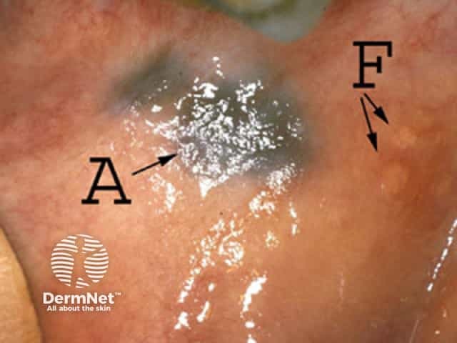
An amalgam tattoo refers to a blue, black or slate grey coloured lesion found in the mouth. An amalgam tattoo most commonly occurs on the gum near an amalgam filling but can occur anywhere in the mouth.
Amalgam is an alloy, or mixture of metals, consisting of mercury, silver, tin and copper. It has been used as a filling material in dentistry for over 150 years[1]. Amalgam fillings are sometimes known as "silver fillings" due to their silver coloured appearance.
Who gets an amalgam tattoo?
People who have amalgam fillings, have had amalgam fillings replaced or who have had a procedure called an apicoectomy (where the tip of the root of a tooth is removed, the area cleaned and a filling is placed in the root), may have an amalgam tattoo.
What causes an amalgam tattoo?
An amalgam tattoo develops when a piece of amalgam filling material becomes inadvertently lodged in the oral mucosa. This can happen whilst the fillings are being placed or polished, or during the removal of a filling following the use of the dental drills.
What are the clinical features of an amalgam tattoo?
An amalgam tattoo presents as a blue, black or slate grey macule on the oral mucosa. They are commonly found on the gum near a tooth with an amalgam filling; however, they have been discovered on the roof of the mouth, inside of the cheeks and on the tongue.

Amalgam tattoo

Amalgam tattoo
How is the diagnosis of an amalgam tattoo made?
An amalgam tattoo can be diagnosed clinically, especially when it is seen adjacent to an amalgam filling. Sometimes an x-ray may be useful, but only if large particles of amalgam are present that will actually show on x-ray. A biopsy may also be used in diagnosing an amalgam tattoo if there is any doubt regarding the exact diagnosis.
What is the treatment for an amalgam tattoo?
As amalgam tattoos are harmless and cause no symptoms, no treatment is required.
References
- Bharti R, Wadhwani KK, Tikku AP, Chandra A. Dental amalgam: An update. J Conserv Dent 2010;13:204–8. PubMed
- Eisen D. Disorders of pigmentation in the oral cavity. Clin Dermatol 2000;18:579–87. PubMed
- Tran HT, Anandasabapathy N, Soldano AC. Amalgam tattoo. Dermatol Online J 2008;14:19. PubMed
- Kauzman A, Pavone M, Blanas N, Bradley G. Pigmented lesions of the oral cavity: review, differential diagnosis, and case presentations. J Can Dent Assoc 2004;70:682–3. PubMed
On DermNet
Other websites
- Disorders of Oral Pigmentation — Medscape Reference
