Main menu
Common skin conditions

NEWS
Join DermNet PRO
Read more
Quick links
Mixed diagnoses – 10 cases (2 of 11)
For each of the ten cases, study the image(s) and then answer the questions. You can click on the image to view a larger version if required.
Each case should take approximately 2 minutes to complete. There is a list of suggested further reading material at the end of the quiz.
When you finish the quiz, you can download a certificate.
Case 1

What is the diagnosis?
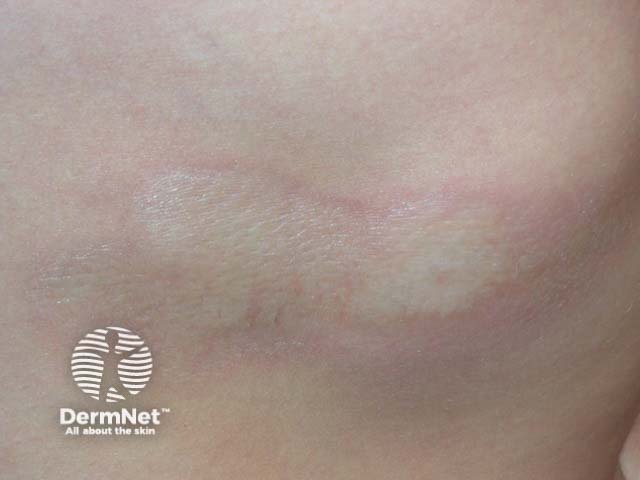
This is plaque-type morphoea, also known as localised scleroderma. It is quite distinct from systemic sclerosis, or generalised scleroderma. Other types of morphoea include linear morphoea (the most common variety in young children), generalized and deep varieties (rare). They are characterized by thickening and induration of the skin and subcutaneous tissue due to excessive collagen deposition. The exact mechanism is unknown but appears to be due to an autoimmune reaction as all types of morphoea are associated with circulating autoantibodies including ANA.
Plaque-type morphoea is characterised by single or few oval indurated plaques one to thirty centimeters in diameter. Initially there is a pink or violaceous macule, which later becomes smooth, ivory white and hairless in the centre. After some months or more often years, the plaque softens leaving an atrophic white or hyperpigmented area.
How is it treated?
No proven effective treatments for morphoea exist. However, some plaques seem to improve with topical calcipotriol and less often with ultrapotent topical steroids. Phototherapy with PUVA or UVA-1 (not available in New Zealand) may be of benefit.
A variety of more toxic agents have been used in severe forms of morphoea, with best results reported from systemic corticosteroids, D-penicillamine, oral vitamin D analogues, cyclosporin, methotrexate and anti-fibrotic agents.
