Main menu
Common skin conditions

NEWS
Join DermNet PRO
Read more
Quick links
Juvenile xanthogranuloma — extra information
Juvenile xanthogranuloma
Authors: Dr Amanda Oakley, Dermatologist, Hamilton, New Zealand; Dr Amy Stanway, Dermatology Registrar, Nottingham, UK. Revised: Dr David Lim, Dermatology Registrar, Auckland, New Zealand. August 2011. Updated: Dr Richa Tripathi, Consultant Dermatologist, Grande International Hospital, Kathmandu, Nepal. Copy edited by Gus Mitchell. October 2021
Introduction Demographics Causes Clinical features Dermoscopy Variation in skin types Complications Diagnosis Differential diagnoses Treatment Outcome
What is juvenile xanthogranuloma?
Juvenile xanthogranuloma is a type of non-Langerhans cell histiocytosis usually limited to the skin in very young children.
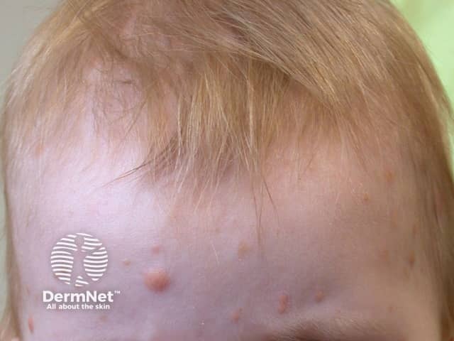

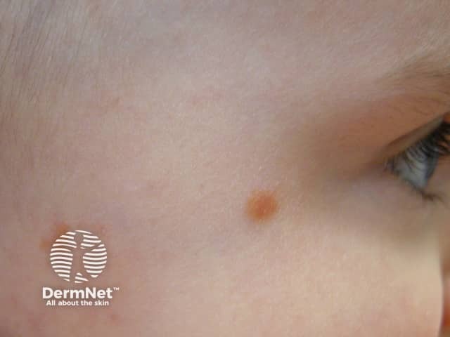
Who gets juvenile xanthogranuloma?
Juvenile xanthogranuloma (JXG) typically presents before one year of age (85%). One third present as a congenital lesion, and adult-onset cases do occur (10%). There is a male predominance in children (up to 7:1) but no difference between the sexes in adult-onset disease. JXG is considered a rare disease in itself, but is the most common type of non-Langerhans histiocytosis.
The incidence of juvenile xanthogranuloma is estimated to be 1 per million in children, however it is probably underdiagnosed.
Up to 10% of patients with neurofibromatosis type I may develop JXG. There have been many single case reports of JXG occurring in association with other conditions.
What causes juvenile xanthogranuloma?
Juvenile xanthogranuloma is a polyclonal proliferation of cholesterol-containing factor XIIIa-positive histiocytes, the cause of which is unknown. Theories include:
- Nonspecific response to injury or viral infection
- Somatic gene mutations
- Activation of extracellular signal-regulated kinase (ERK).
What are the clinical features of juvenile xanthogranuloma?
- Solitary asymptomatic skin lesion (80%)
- Multiple lesions and systemic involvement can occur
- Commonest site — head and neck
- Less common sites
- Trunk and limbs
- Rare mucocutaneous sites include hands and feet, anogenital, oral cavity
- Eye is the most common extracutaneous site (0.5%); unilateral involvement of the iris; typically presents as a red eye, sometimes without skin involvement
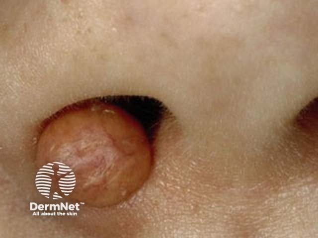
- Papules and nodules, 1–10 mm in diameter, smooth and firm; large lesions can ulcerate
- Lesion colour progresses from red through yellow to brown before fading
- Atypical forms – include giant, generalised, clustered, subcutaneous
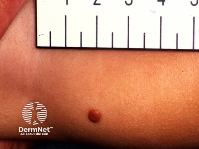
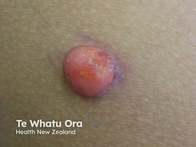
Dermoscopy of juvenile xanthogranuloma
- Setting sun pattern
- Central yellow-orange
- Pale yellow clouds
- Branched linear vessels
How do clinical features vary in differing types of skin?
Juvenile xanthogranuloma is probably underdiagnosed in dark skin as erythema can be difficult to appreciate and the asymptomatic nature and self-limiting course mean a solitary lesion may not be noticed.
What are the complications of juvenile xanthogranuloma?
- Ulceration and atrophic scarring, particularly of a congenital lesion
- Ocular lesions can lead to hyphema, glaucoma, and/or loss of vision
- Systemic involvement including liver and bone – often, but not always, associated with multiple skin lesions
- Multiorgan involvement can be fatal (rare)
How is juvenile xanthogranuloma diagnosed?
Juvenile xanthogranuloma is rarely a clinical diagnosis, although dermoscopy is characteristic. Confirmation may require biopsy [see Juvenile xanthogranuloma pathology].
Patients with more than two skin lesions, early onset lesions, and/or a unilateral red eye or ocular symptoms should be referred for an eye examination.
Following a full skin examination, a typical solitary lesion may be investigated only if clinically indicated. If two or more skin lesions are identified investigations should be directed by clinical symptoms and signs, and may include:
- Blood tests — full blood count (bone marrow biopsy if cytopenic), liver and kidney function tests
- Ultrasound — of abdomen and lymph nodes
- Radiology — X-rays, brain MRI, and/or CT scan.
What is the differential diagnosis for juvenile xanthogranuloma?
- Solitary lesion — mastocytoma, benign cephalic histiocytosis, necrobiotic xanthogranuloma
- Multiple lesions — molluscum contagiosum, Langerhans cell histiocytosis, multicentric reticulohistiocytosis
What is the treatment for juvenile xanthogranuloma?
- Solitary lesion
- Observation if asymptomatic
- Simple excision if causing functional impairment or psychosocial effects
- Eye involvement — topical or intralesional steroids
- Systemic disease
- Observation
- Chemotherapy for organ dysfunction
What is the outcome for juvenile xanthogranuloma?
Juvenile xanthogranuloma is usually self-limiting with skin lesions regressing in 3–6 years. Even systemic JXG resolves spontaneously in most cases. Skin lesions of juvenile xanthogranuloma in adults tend to be more persistent than JXG in children.
Bibliography
- Hernández-San Martín MJ, Vargas-Mora P, Aranibar L. Juvenile xanthogranuloma: an entity with a wide clinical spectrum. Actas Dermosifiliogr (Engl Ed). 2020;111(9):725–33. doi:10.1016/j.ad.2020.07.004. Journal
- Höck M, Zelger B, Schweigmann G, et al. The various clinical spectra of juvenile xanthogranuloma: imaging for two case reports and review of the literature. BMC Pediatr. 2019;19(1):128. doi:10.1186/s12887-019-1490-y. Journal
- Oza VS, Stringer T, Campbell C, et al. Congenital-type juvenile xanthogranuloma: a case series and literature review. Pediatr Dermatol. 2018;35(5):582–7. doi:10.1111/pde.13544. PubMed
- So N, Liu R, Hogeling M. Juvenile xanthogranulomas: examining single, multiple, and extracutaneous presentations. Pediatr Dermatol. 2020;37(4):637–44. doi:10.1111/pde.14174. PubMed
- Xu J, Ma L. Dermoscopic patterns in juvenile xanthogranuloma based on the histological classification. Front Med (Lausanne). 2021;7:618946. doi:10.3389/fmed.2020.618946. Journal
On DermNet
- Histiocytosis
- Intralymphatic histiocytosis
- Langerhans cell histiocytosis
- Malignant histiocytosis
- Reticulohistiocytosis
- Xanthoma
Other websites
- Dermatologic manifestations of juvenile xanthogranuloma — Medscape
- Juvenile xanthogranuloma — Medscape
