Main menu
Common skin conditions

NEWS
Join DermNet PRO
Read more
Quick links
Acral lentiginous melanoma images
Go to the acral lentiginous melanoma topic page
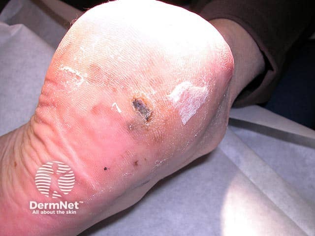
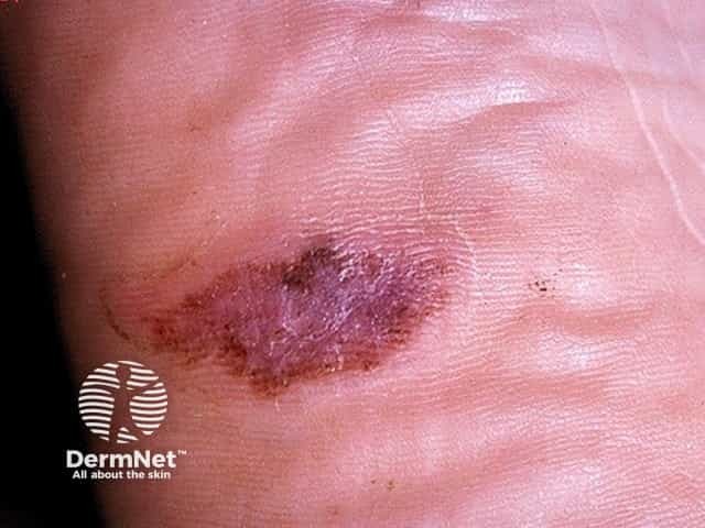
An irregularly pigmented lesion on the heel - histology showed a thin acral lentiginous melanoma
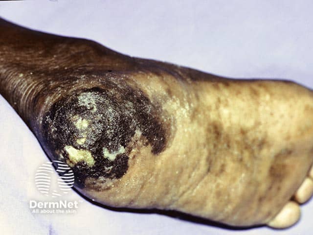
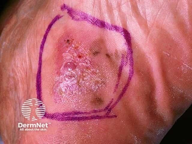
An advanced acral lentiginous melanoma in skin of colour on the heel
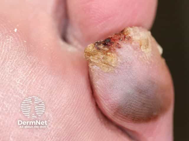
An irregular grey pigmented lesion on the toe that has become both hyperkeratotic and ulcerated on the toe tip
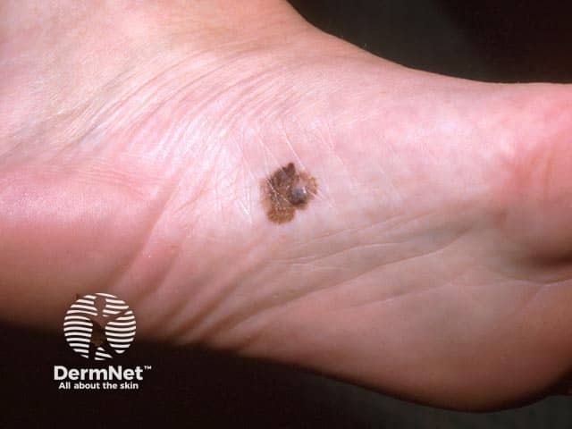
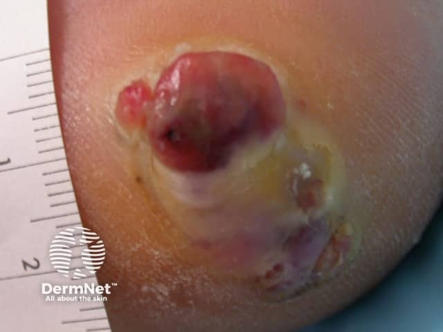
A nodule arrising within an acral lentiginous melanoma as it enters a vertical growth phase
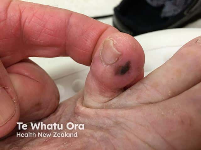
Melanoma in situ on the toe - acral lentiginous melanoma type (ALM-patient1)
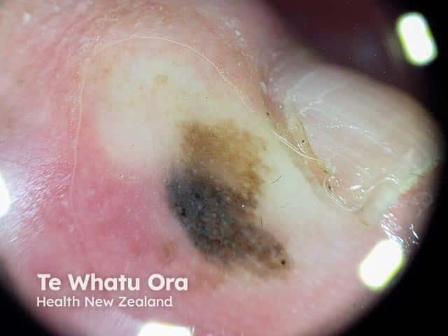
Dermoscopy of an acral melanoma in situ on the toe (ALM-patient1)
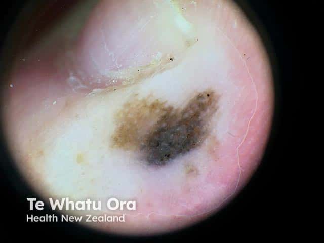
Dermoscopy of an acral melanoma in situ on the toe (ALM-patient1)
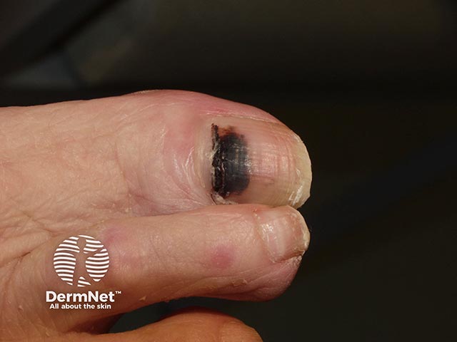
Brown-black irregularly-marginated pigment with extension into the cuticle - a subungual melanoma
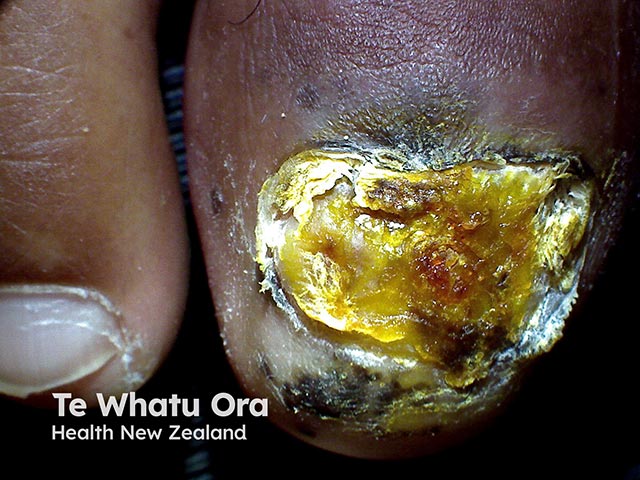
Complete destruction of the nail plate with deep pigmentation into the digit pulp and the proximal nail fold - a subungal melanoma (ALM-patient2)
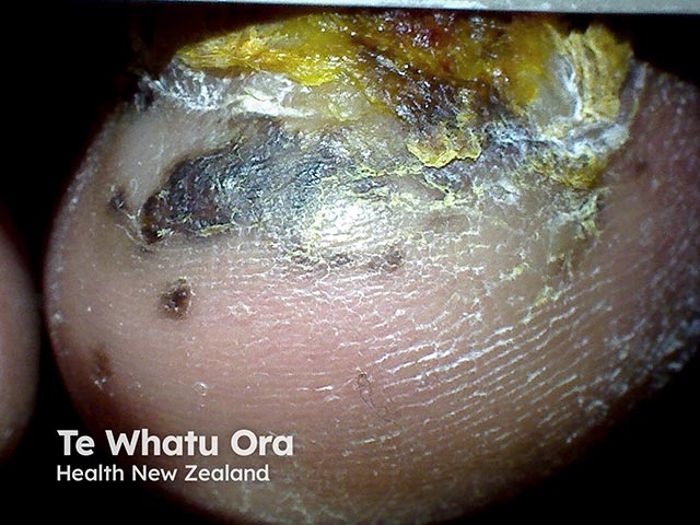
Irregular pigmentation of the end of the great toe and destruction of the nail plate due to a subungual melanoma (ALM-patient2)
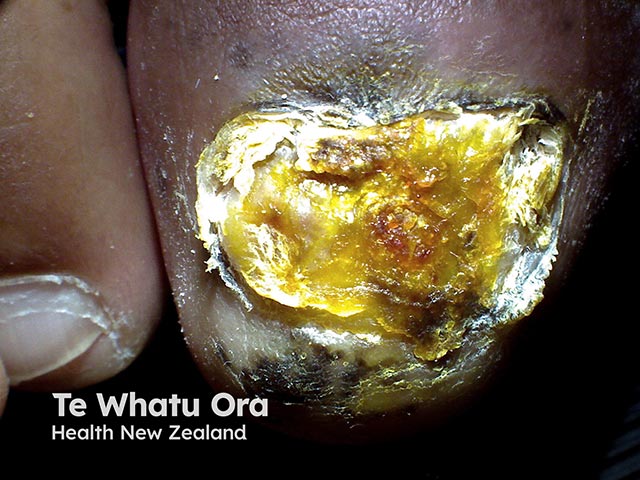
Complete destruction of the nail plate with deep pigmentation into the digit pulp and the proximal nail fold. - a subungual melanoma (ALM-patient2)
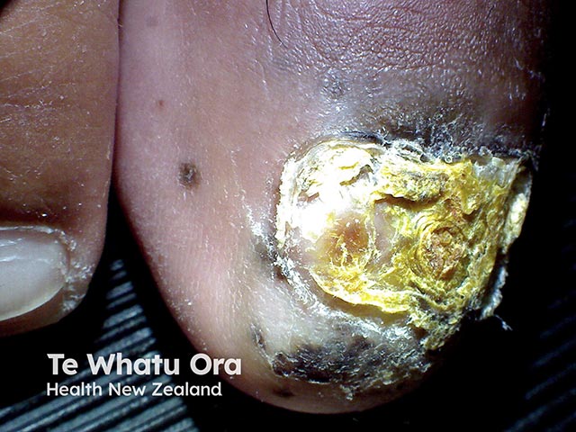
Complete destruction of the nail plate with pigmentation extending into the digit pulp and the proximal nail fold - a subungual melanoma (ALM-patient2)
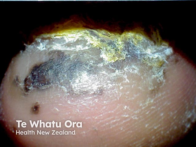
Irregular pigmentation of the hyponychium due to a subungual melanoma (ALM-patient2)
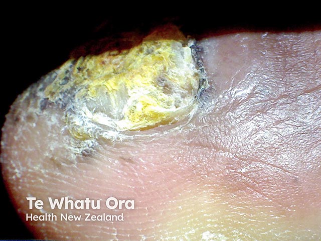
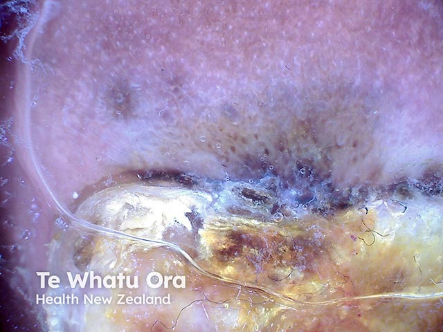
Dermoscopic image of a subungual melanoma of the great toenail - 0.5 mm in depth (ALM-patient2)
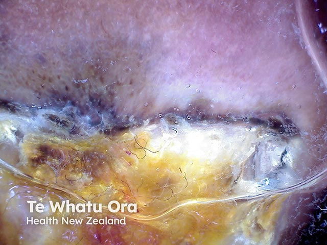
Dermoscopic image of a subungual melanoma of the great toenail - 0.5 mm in depth (ALM-patient2)
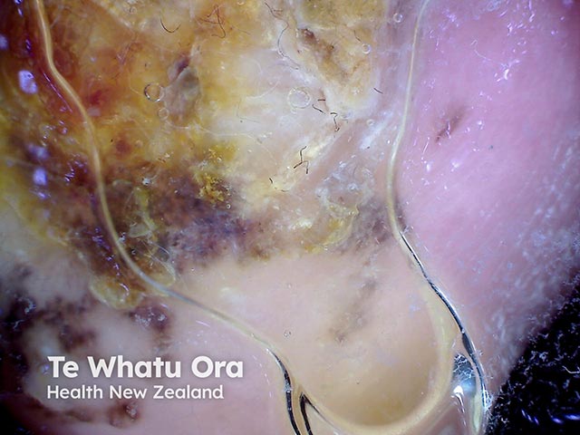
Dermoscopic image of a subungual melanoma of the great toenail - 0.5 mm in depth (ALM-patient2)
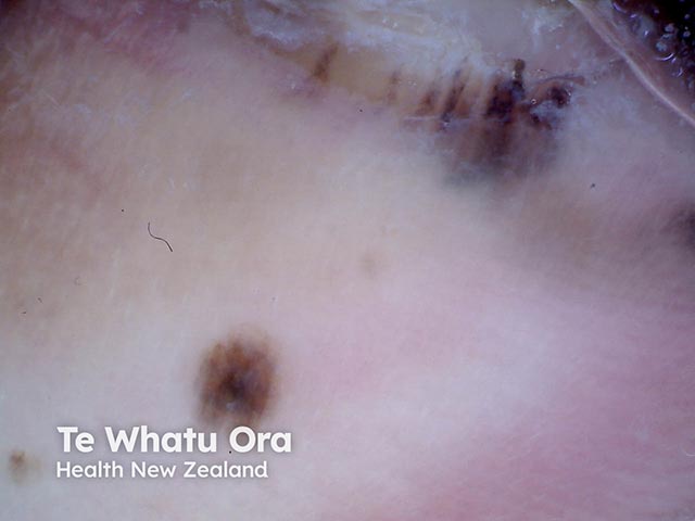
Pigmentation proximal to the nail fold due to a subungual melanoma (ALM-patient2)
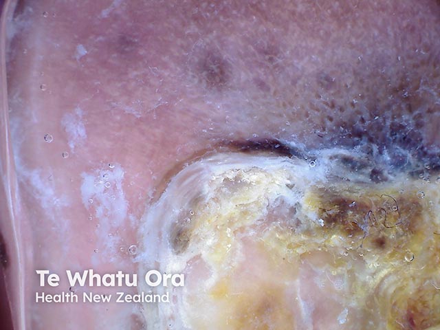
Melanoma of the nail unit - dermoscopy shows pigmentation of the proximal nail fold (Hutchinson's sign) (ALM-patient2)
Go to the acral lentiginous melanoma topic page
Acral lentiginous melanoma images
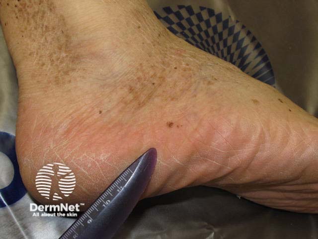
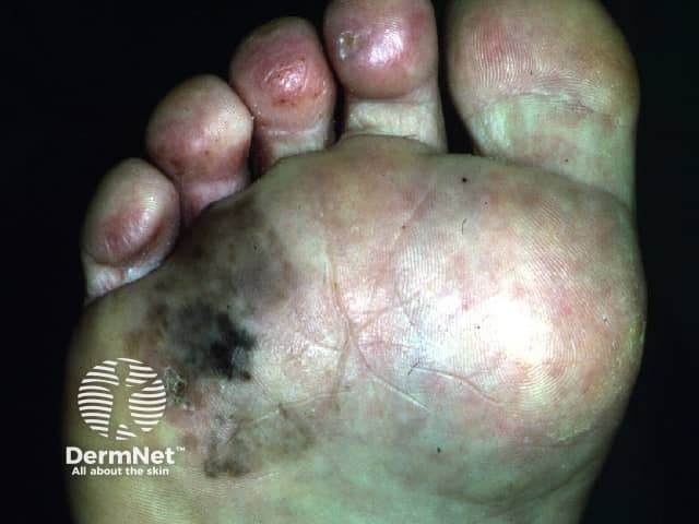
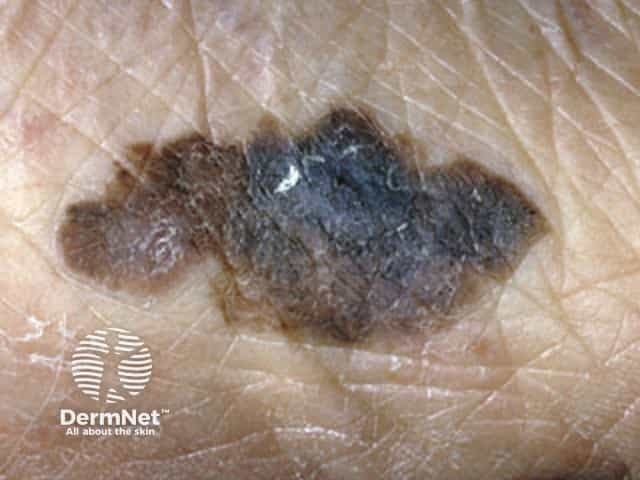
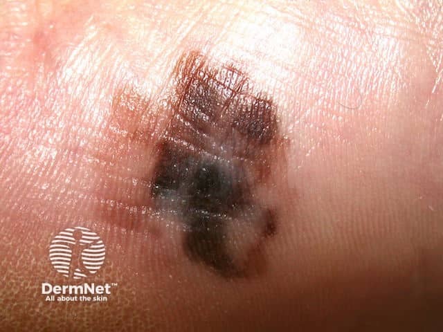
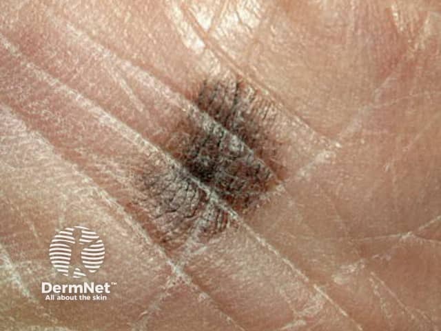
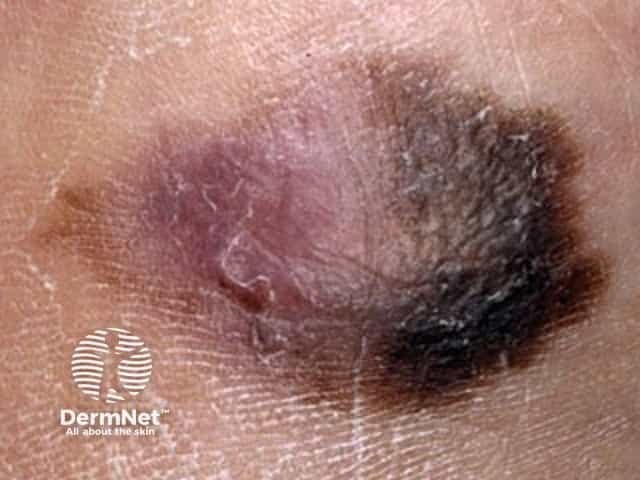
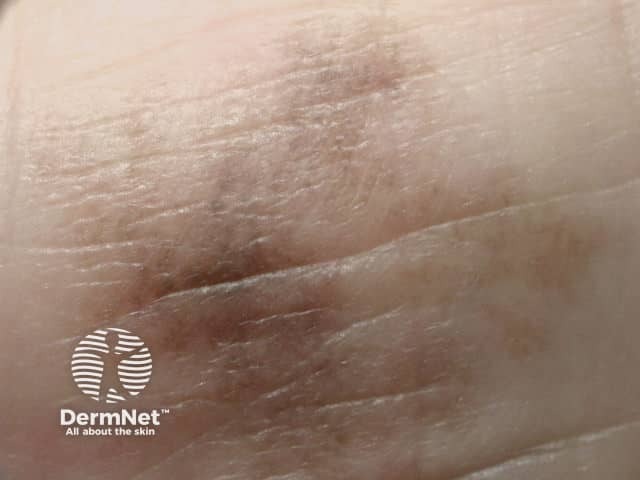
Irregular margin and pigmentation of an acral lentiginous melanoma
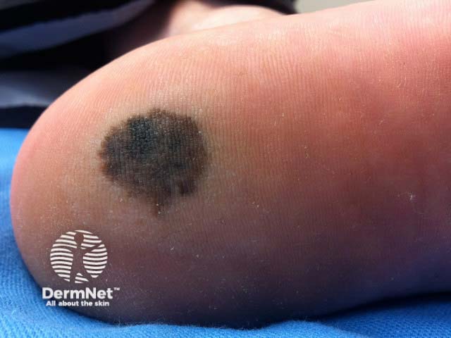
Slowly enlarging irregularly pigmented lesion on the heel present for 3 years
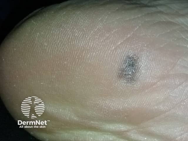
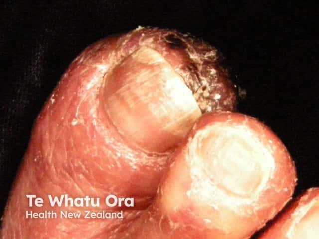
Right Foot Inner
Go to the acral lentiginous melanoma topic page
Acral lentiginous melanoma dermoscopy images
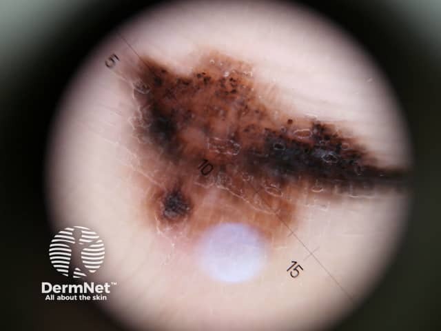
Asymmetrical structure and colour with multicomponent pattern
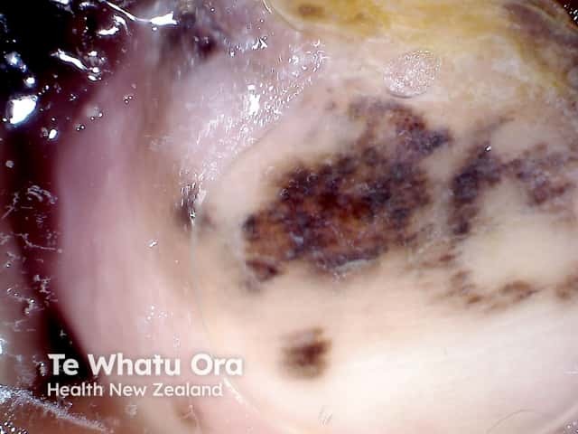
Asymmetrical structure and colour with multicomponent pattern
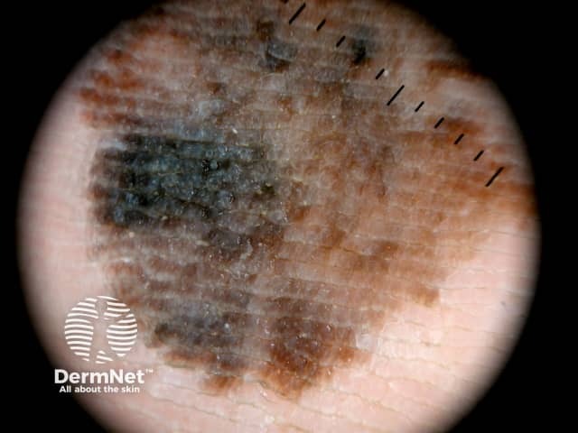
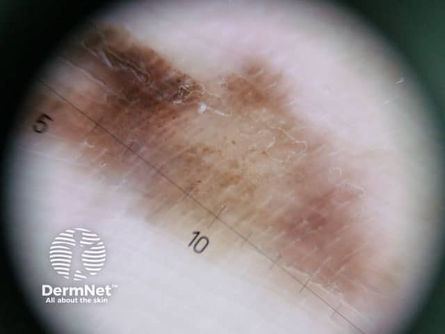
Parallel ridge pattern, asymmetrical structure and diffuse pigmentation
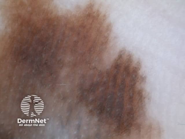
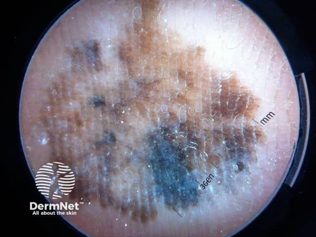
Dermoscopy of an acral lentiginous melanoma showing variable pigmentation and areas of regression
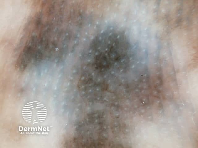
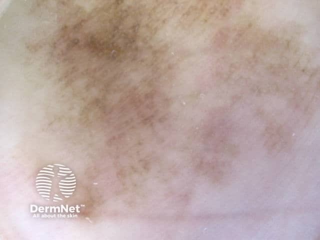
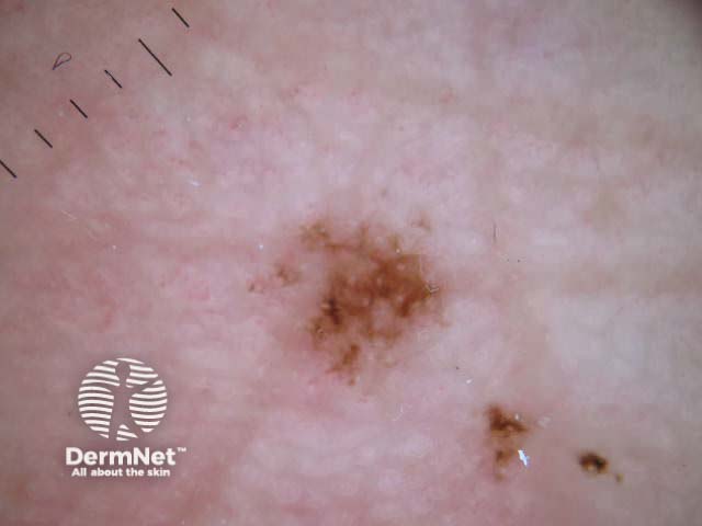
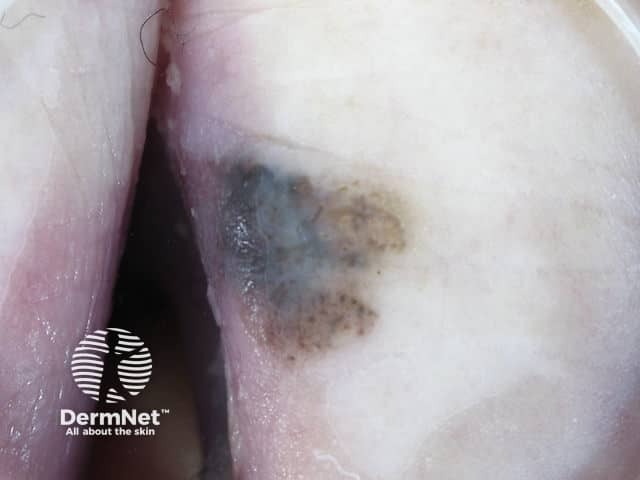
Dorsum 3rd Toe Right Foot
Go to the acral lentiginous melanoma topic page
Acral lentiginous melanoma macro and dermoscopy image pairs
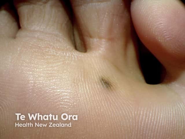
Acral lentiginous melanoma 1 macro

Acral lentiginous melanoma 1 dermoscopy
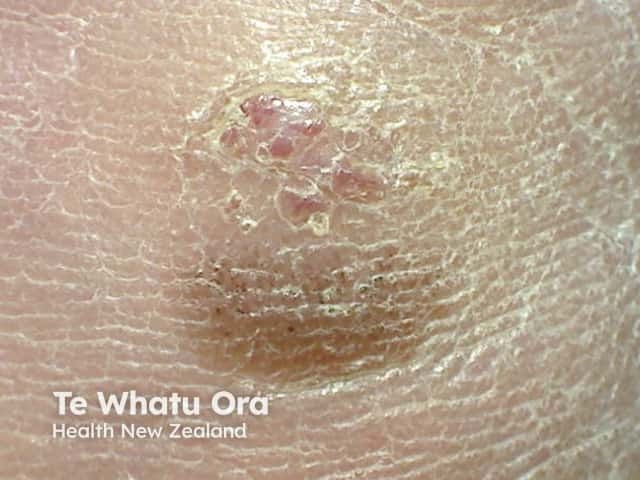
Acral lentiginous melanoma 2 macro
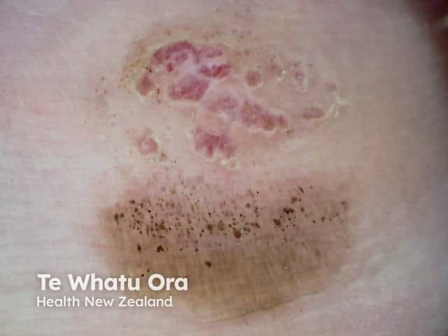
Acral lentiginous melanoma 2 dermoscopy
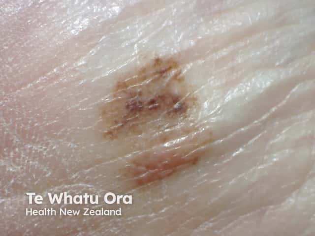
Acral lentiginous melanoma 3 macro
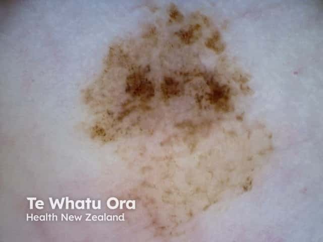
Acral lentiginous melanoma 3 dermoscopy
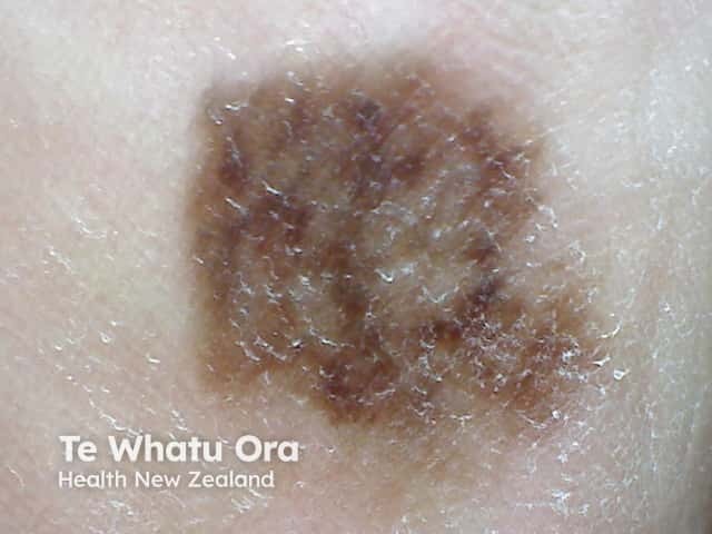
Acral lentiginous melanoma 4 macro
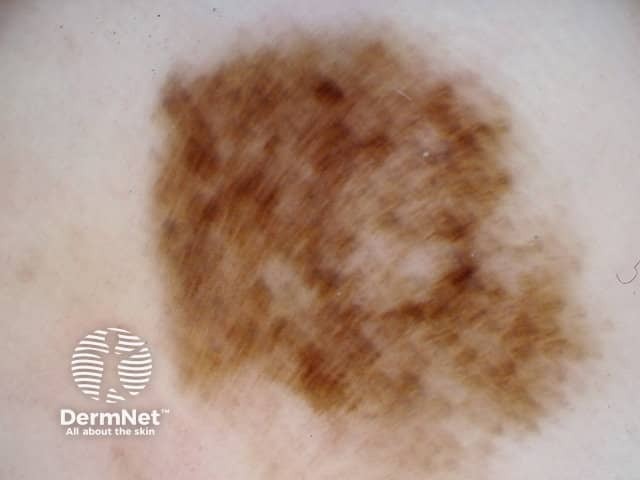
Acral lentiginous melanoma 4 dermoscopy
