Main menu
Common skin conditions

NEWS
Join DermNet PRO
Read more
Quick links
Dermoscopy of basal cell carcinoma
Created 2008.
Learning objectives
- Describe dermoscopic features of basal cell carcinoma
Introduction
Dermoscopy is useful to distinguish pigmented basal cell carcinoma from other pigmented lesions. There are specific features that help to distinguish these. Pigment may be grey, brown, blue or black. They are rarely completely pigmented in white-skinned individuals.
Pigmented basal cell carcinoma
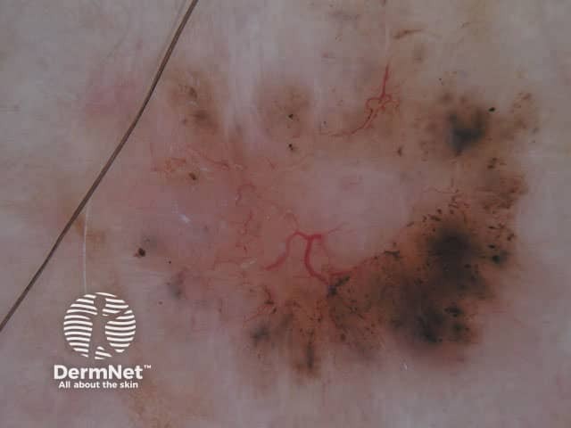
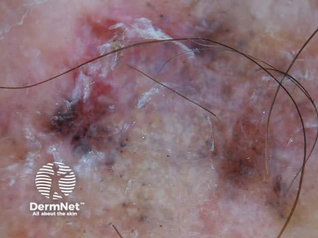
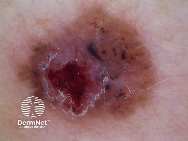
The dermoscopic features of pigmented basal cell carcinoma include:
- Absence of pigment network
- Linear and arborising (branch-like) telangiectasia
- Structureless or leaf-like areas on the periphery of the lesion
- Large blue-grey ovoid nests or blotches
- Multiple blue-grey globules
- Specks of brown and grey pigment
- Spoke wheel areas (radial projections from a well circumscribed dark central hub)
- Focal ulceration
Blue ovoid nests Arborising blood vessels Focal ulceration Ulceration Spoke-wheel pigmentation Flecks of pigment Arborising telangiectasia Flecks of pigment Flecks of pigment Deeply pigmented border Multicoloured lesion Large nodule 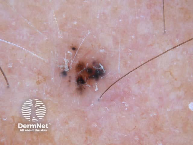
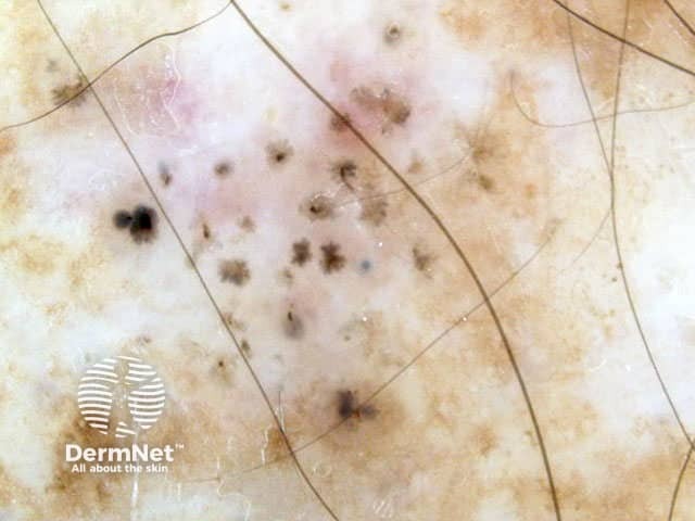
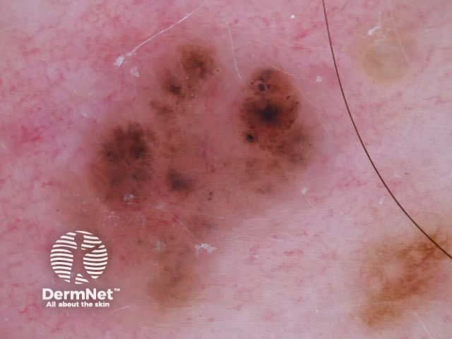
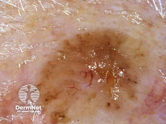
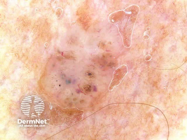
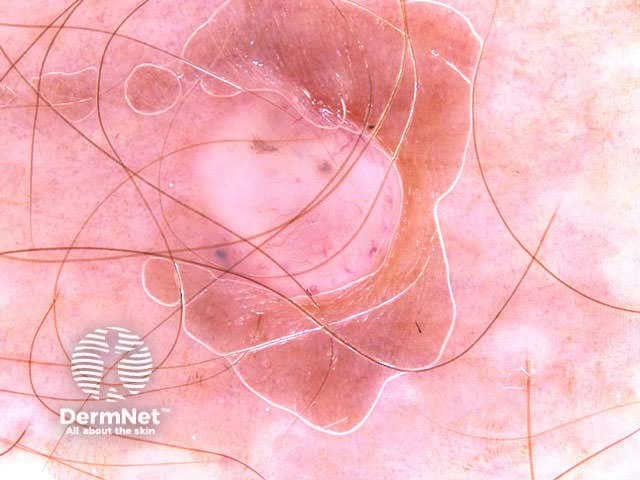
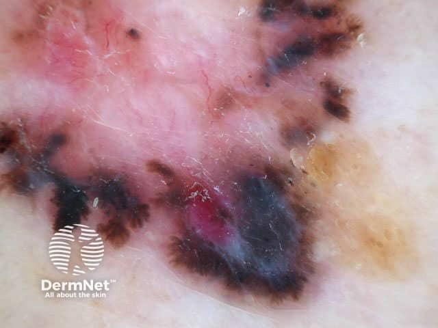
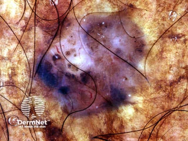
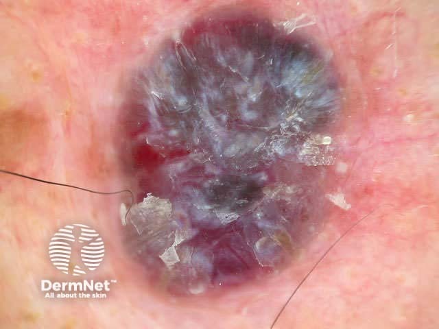
In some cases, it may be difficult to distinguish deeply pigmented or even non-pigmented basal cell carcinoma from melanoma.
Non-pigmented basal cell carcinoma
Non-pigmented basal cell carcinomas are much more common than pigmented basal cell carcinoma. They contain flecks of grey colour.
Experienced dermoscopists can often diagnose superficial basal cell carcinomas by their typical bluish-pink colour, asymmetrical arborising vessels and focal ulceration. Slight scaling and white areas of regression may also be present. Chrystalline structures, i.e. white shiny lines, strands and larger irregular-shaped white areas, are common in all histological types of basal cell carcinoma. These short parallel or disordered lines and roundish white structures are often only visible on polarised dermoscopy.
Nodular basal cell carcinomas lose the blue hue and instead have a white rim around central ulceration. Milia may be present. Disordered and streaky crystalline structures may be seen.
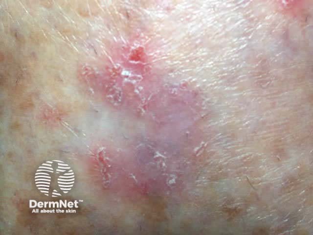
Bluish, scale, ulceration
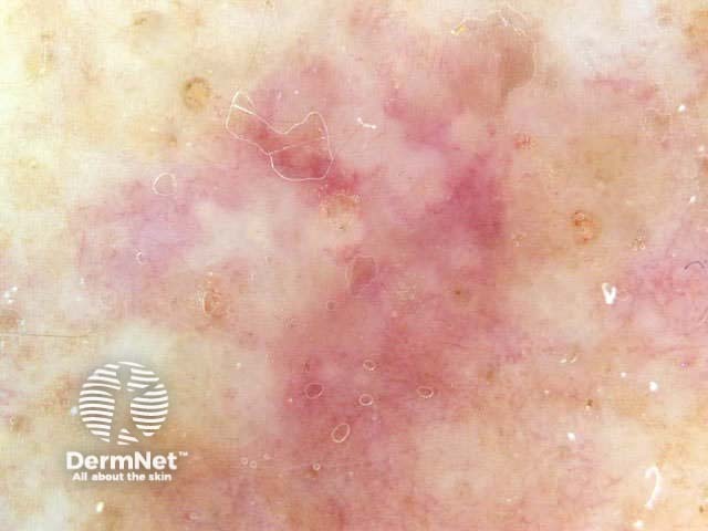
Subtle blue colour
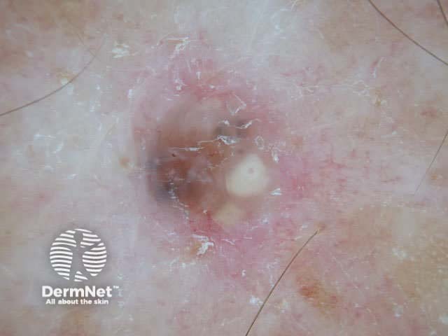
Milia formation
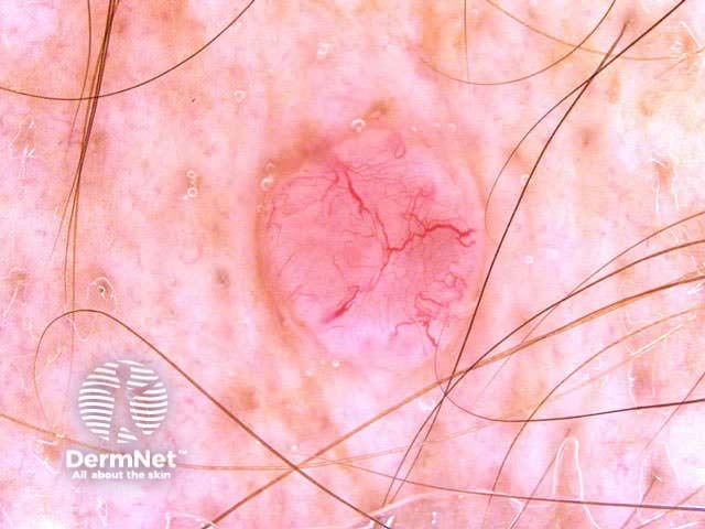
Prominent vasculature
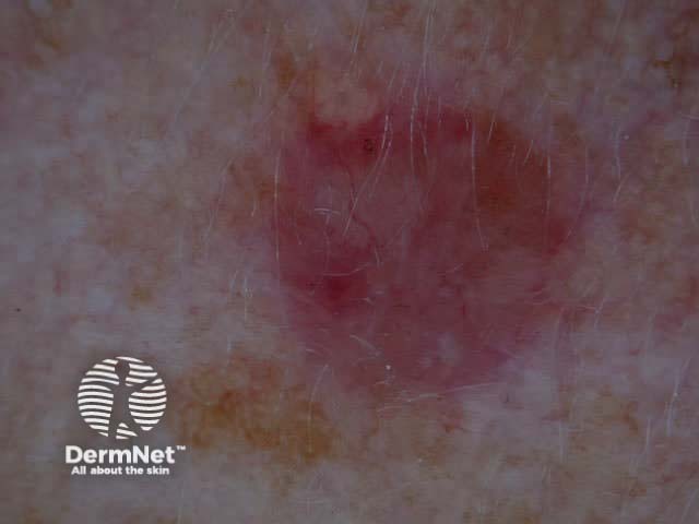
Vascular bcc
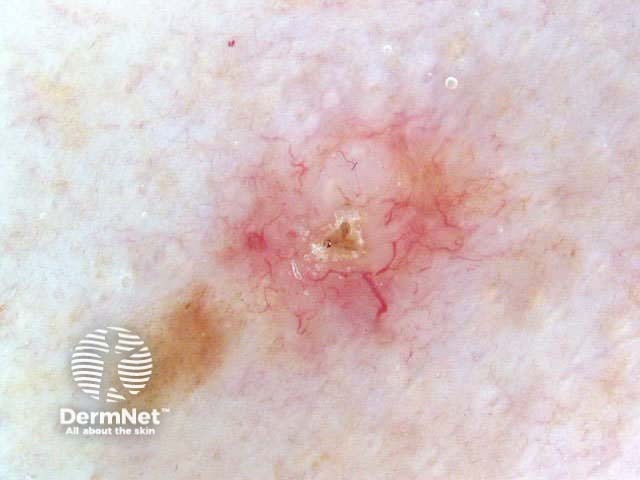
Crusting, telangiectasia
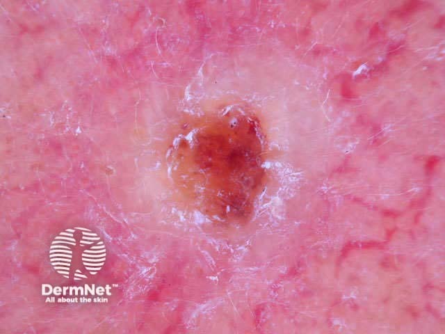
White nodule, telangiectasia
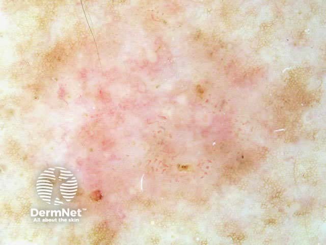
Tiny ulcers
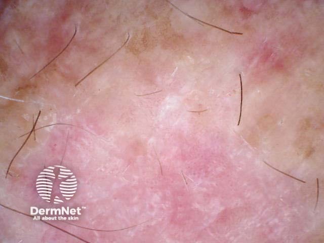
Superficial basal cell carcinoma
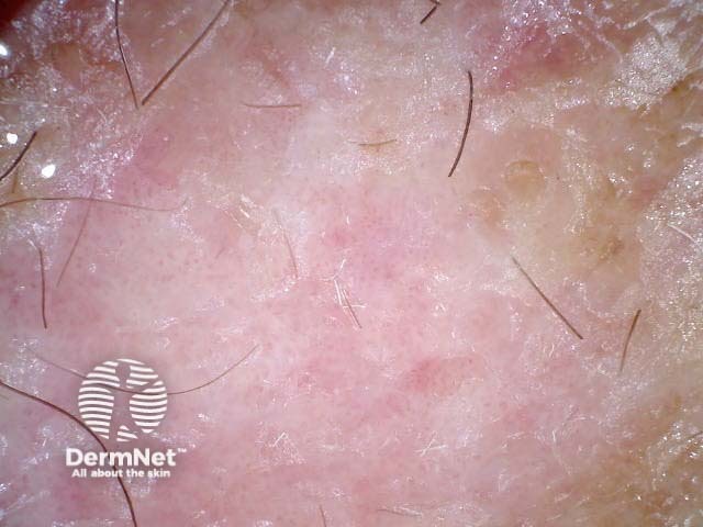
Superficial basal cell carcinoma
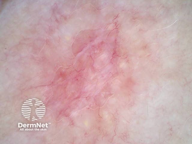
Cystic basal cell carcinoma (polarised view showing crystalline structures)
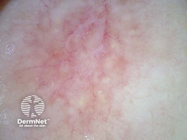
Cystic basal cell carcinoma (non-polarised view)
Activity
Imiquimod cream has restricted subsidy by PHARMAC for treating certain superficial basal cell carcinomas (New Zealand, September 2008). Observe the inflammatory response to treatment using dermoscopy.
References
- Menzies SW. Dermoscopy of pigmented basal cell carcinoma. Clin Dermatol. 2002 May-Jun;20(3):268-9. Medline.
- Menzies SW, Westerhoff K, Rabinovitz H, Kopf AW, McCarthy WH, Katz B. Surface microscopy of pigmented basal cell carcinoma. Arch Dermatol. 2000 Aug;136(8):1012-6. Medline.
- Kreusch JF. Vascular patterns in skin tumors. Clin Dermatol. 2002 May-Jun;20(3):248-54. Medline.
On DermNet
- Basal cell carcinoma
- Imiquimod
- Dermatoscope overview
- Dermatoscopy quizzes
- Dermatoscopy Information for patients
Books about skin diseases
See the DermNet bookstore.
