Main menu
Common skin conditions

NEWS
Join DermNet PRO
Read more
Quick links
An Afghan female with painful ulcers
Last reviewed: May 2023
Author: Dr Emily Balfour, Senior House Officer; Dr Burcu Isler, Infectious Diseases Consultant, Princess Alexandra Hospital, Australia (May 2023). Reviewing dermatologist: Dr Ian Coulson
Edited by the DermNet content department


Background
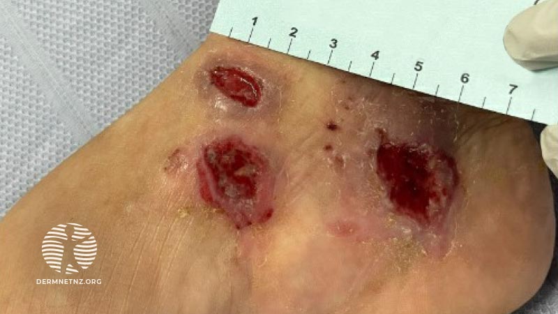
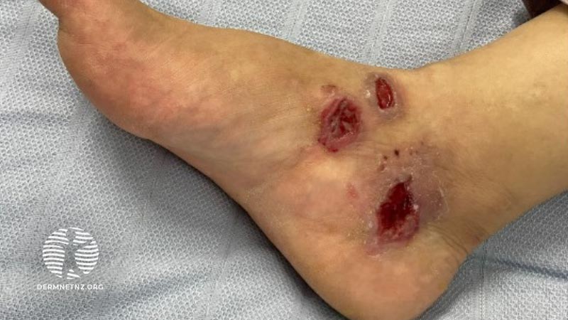
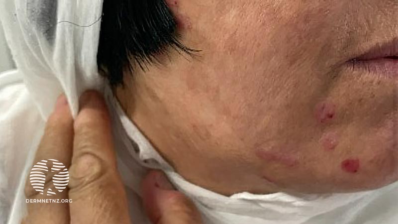
A 50-year-old Afghan female presents with a 3-month history of progressive, painful ulcers overlying the right medial malleolus, right dorsal hand, and right lateral chin. There was no mucosal involvement.
She had a history of cutaneous leishmaniasis, with similarly distributed lesions. These were successfully treated in Afghanistan some years ago. She migrated to Australia 6 months ago and has been otherwise well since.
List 4 differential diagnoses
- Cutaneous leishmaniasis — the patient is from an endemic region and has a positive past history. Leishmaniasis recividans (LR) should be considered given that there is facial involvement and multiple lesions distributed over previously healed lesions.
- Anthrax — endemic to Afghanistan and cutaneous anthrax is the most common presentation.
- Lupus vulgaris — chronic direct infection of the skin with tuberculosis.
- Atypical mycobacterial infection — M. fortuitum and M. chelonae are both found in Afghanistan and may cause skin ulcers.
What is the most likely subspecies, and what might a punch biopsy show?
L. tropica is the most common isolate in Afghanistan and it causes primarily cutaneous disease. Histology shows numerous macrophages with intracellular organisms, compatible with leishmania amastigotes. Additional PCR is important to ascertain the causative species because clinical sequelae vary within the leishmania class.
Culture and PCR confirmed leishmaniasis tropica.
Additional PCR is important given that leishmaniasis recividans is a differential and it demonstrates a scarcity of amastigotes, easily leading to a misdiagnosis as lupus vulgaris.
How is this treated?
Clinical observation is reasonable for immunocompetent patients with uncomplicated lesions attributable to a leishmania species not associated with increased risk of mucosal lesions.
In this case, treatment is indicated because as lesions have not spontaneously regressed over three months. First-line therapy is topical paromomycin. Because this case was diagnosed in Brisbane, Australia, we were unable to access this first-line therapy. Instead, a 7 day course of intravenous amphotericin was administered.
