Main menu
Common skin conditions

NEWS
Join DermNet PRO
Read more
Quick links
Granular cell tumour — extra information
Granular cell tumour
Last reviewed: May 2023
Authors: Dr Vera Miao, Northern Sydney; and Clinical Professor Saxon D Smith, Dermatologist, Sydney Adventist Hospital Clinical School, Australia (2023)
Previous contributors: Dr Todd Gunson, Dermatology Registrar (2008)
Reviewing dermatologist: Dr Ian Coulson
Edited by the DermNet content department
Introduction Demographics Causes Clinical features Variation of skin types Complications Diagnosis Differential diagnoses Treatment Outcome
What is a granular cell tumour?
Granular cell tumours are rare, generally benign, soft tissue neoplasms believed to originate from Schwann cells (cells that provide myelin insulation to nerves). They can occur in the skin, tongue, breast, gastrointestinal tract, and respiratory tract.
Granular cell tumours, also known as a granular cell myoblastoma or Abrikossoff tumour, display characteristic pathologic findings under the microscope. The vast majority of granular cell tumours are benign lesions; less than 2% are malignant.

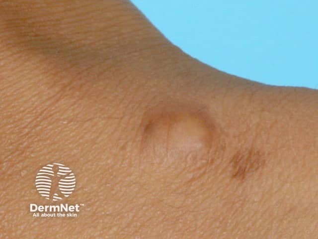
Granular cell tumour on the wrist - the clinical diagnosis is seldom considered, and relies on histopathological assessment

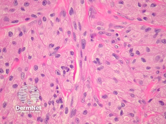
High power histopathology - demonstrates fine granularity of the tumour cytoplasm
Who gets granular cell tumours?
Granular cell tumours are rare and can occur in anyone. They tend to be more common in females and dark-skinned people. Most develop between the fourth to sixth decades of life.
What causes granular cell tumours?
Certain genetic mutations have been linked to granular cell tumours, although the exact mechanisms remain poorly understood.
In some case reports, multiple granular cell tumours have been associated with certain rare syndromes, including Leopard syndrome, Noonan syndrome, and neurofibromatosis type I.
What are the clinical features of granular cell tumour?
Granular cell tumours may occur at any body site and are most commonly reported in the skin, tongue, breast, gastrointestinal tract, and respiratory tract. They occur in the skin or subcutaneous tissue in 30–40% of cases, most frequently on the head and neck.
Clinical features of a granular cell tumour include:
- A solitary, small (usually 1–3 cm) nodule
- Skin-coloured or brown-red
- Smooth or slightly rough surface
- Slow-growing
- Most often painless; can occasionally be mildly itchy or tender.
Multiple granular cell tumours may occur in up to 25% of cases.
Clinical features of a granular cell tumour that may suggest malignancy include:
- Size >4 cm
- Rapid growth
- Ulceration or necrosis
- Lymph node involvement.
How do clinical features vary in differing types of skin?
Multiple granular cell tumours are more common in dark-skinned people. Lesions may vary in colour and have been described as white, grey, brown, or tan.
What are the complications of granular cell tumour?
- Recurrence: occurs in 2–8% of cases even when surgical resection margins are clear; recurrence rates are higher with malignant granular cell tumours.
- Wound infection following excision.
- Rarely, granular cell tumours can be malignant lesions.
- Regional or distant metastases.
How is granular cell tumour diagnosed?
Diagnosis is made by skin biopsy. Excision biopsy is preferred to punch or shave biopsy. Histological features include characteristic granules within the cytoplasm of large tumour cells.
What is the differential diagnosis for granular cell tumour?
What is the treatment for granular cell tumour?
- Complete surgical excision with clear margins.
- Mohs micrographic surgery may be appropriate for tumours located in cosmetically and functionally important locations.
- Sentinel lymph node biopsy may be recommended for lesions determined to be malignant.
- Continued monitoring after excision for signs of recurrence or metastases.
What is the outcome for granular cell tumour?
- Benign: prognosis is excellent with complete surgical excision.
- Malignant: 74% survival rate at 5 years and 65% survival rate at 10 years.
Bibliography
- Amphlett A. An update on cutaneous granular cell tumours for dermatologists and dermatopathologists. Clin Exp Dermatol. 2022;47(11):1916–22. doi: 10.1111/ced.15309. Journal
- Aoyama K, Kamio T, Hirano A, et al. Granular cell tumors: a report of six cases. World J Surg Oncol. 2012;10:1–6. doi: 10.1186/1477-7819-10-204. Journal
- Fanburg-Smith JC, Meis-Kindblom JM, Fante R, Kindblom LG. Malignant granular cell tumor of soft tissue: Diagnostic criteria and clinicopathologic correlation. Am J Surg Pathol. 1998;22(7):779–94. doi: 10.1097/00000478-199807000-00001. Journal
- Singh VA, Gunasagaran J, Pailoor J. Granular cell tumour: malignant or benign? Singapore Med J. 2015;56(9):513. doi: 10.11622/smedj.2015136. Journal
- Yilmaz AD, Unlu RE, Orbay H, Sensoz O. Recurrent granular cell tumor: how to treat. J Craniofac Surg. 2007;18(5):1187–9. doi: 10.1097/scs.0b013e3181572663. Journal
- Pressoir R, Chung EB. Granular cell tumor in black patients. J Natl Med Assoc. 1980;72(12):1171–5. PubMed
On DermNet
Other websites
- Granular cell tumours — MedScape
