Main menu
Common skin conditions

NEWS
Join DermNet PRO
Read more
Quick links
Lichen myxoedematosus — extra information
Lichen myxoedematosus
Author: Dr Julie Smith MBChB, Dept of Dermatology, Greenlane Hospital, Auckland, New Zealand, 2005. Updated by Dr Eugene Tan, Dept of Dermatology, Waikato Hospital, Hamilton, New Zealand, 2008.
Introduction Causes Scleromyxoedema Localised lichen myxoedematosus Investigations Treatment Nephrogenic fibrosing dermopathy
What is lichen myxoedematosus?
Lichen myxoedematosus (lichen myxedematosus) is also known as papular mucinosis. It is a rare skin disorder characterised by deposits of mucin in the skin.
Lichen myxoedematosus has localised and generalised forms. The localised form has a more favourable course compared to the generalised form, scleromyxoedema (scleromyxedema), which can involve other organs and is sometimes fatal.
Although the term scleromyxoedema and lichen myxoedematosus have often been used interchangeably, scleromyxoedema refers to the generalised form.
Classification of lichen myxoedematosus (LM)
Generalised form:
- Scleromyxoedema
Localised forms:
- Discrete papular LM
- Acral persistent papular mucinosis
- Juvenile and adult variants of self-healing papular mucinosis
- Papular mucinosis of infancy
- Nodular LM.
Atypical forms:
- Scleromyxoedema without monoclonal gammopathy
- Localised LM with monoclonal gammopathy
- Localised LM with mixed features of different subtypes
- Not otherwise specified.
What causes lichen myxoedematosus?
No specific cause has been found for lichen myxoedematosus.
Scleromyxoedema is nearly always associated with monoclonal gammopathy, in which abnormally high levels of an immunoglobulin called a paraprotein are found in the blood. Usually these are IgG-lambda light chain molecules. A small increase in the number of plasma cells in the bone marrow may be found.
In other cases, scleromyxoedema has been reported in association with cancers of the bone marrow such as myeloma, lymphoma and leukaemia.
A few cases of localised lichen myxoedematosus have been linked with HIV infection, Hepatitis C virus, exposure to toxic oil and contaminated L-tryptophan.
Scleromyxoedema
Scleromyxoedema often affects adults of 30–50 years of age. Men and women are equally affected.
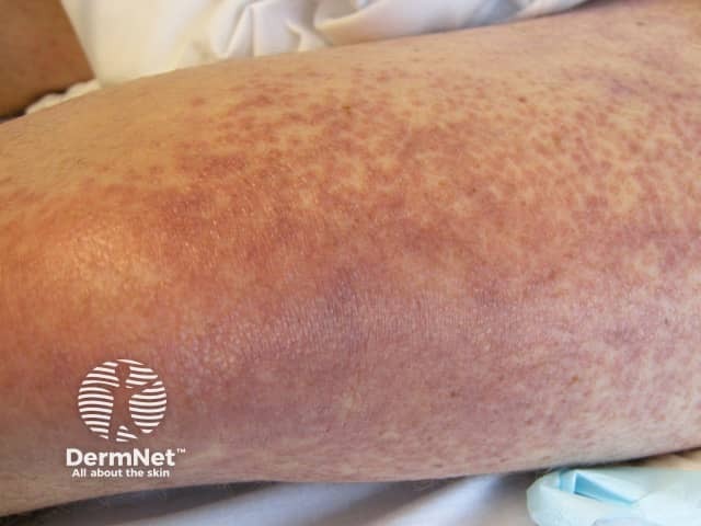
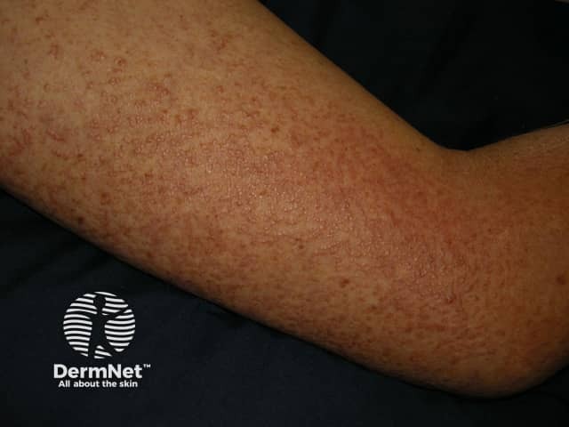
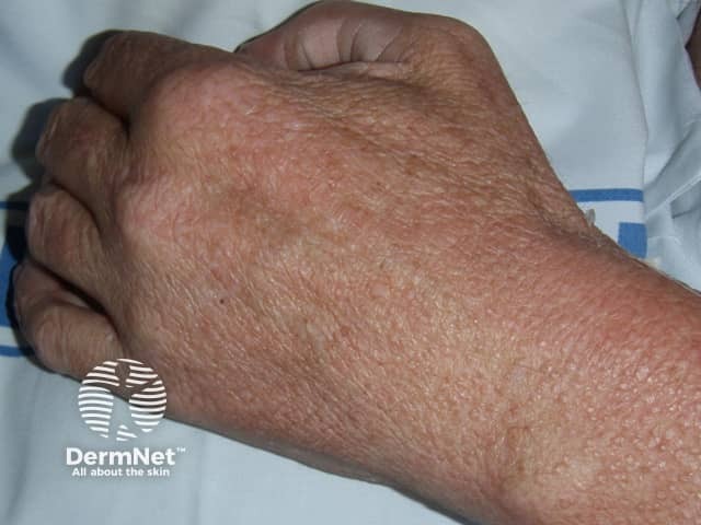
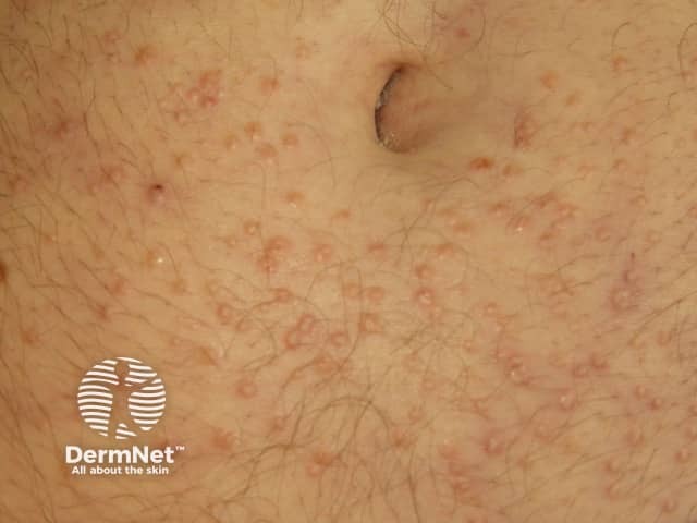
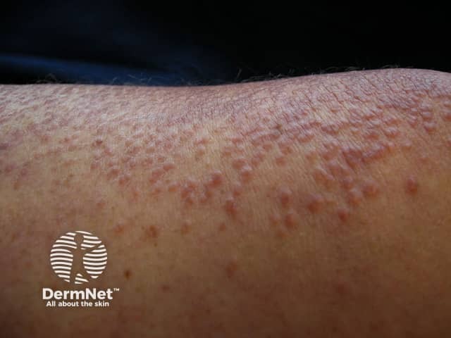
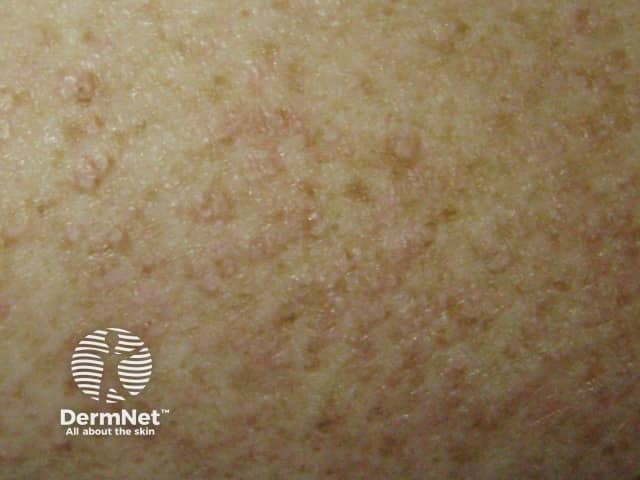
Scleromyxoedema is characterised by widespread skin-coloured papules on the face, trunk and extremities. Mucous membranes and the scalp are not involved.
The papules (small bumps) are 2–3 mm in diameter, waxy, and closely arranged in lines. They may become red-brown in colour.
As the condition progresses, deep furrows may develop across the brow. Skin stiffening of the skin, fingers (sclerodactyly) and reduced mobility of the mouth may occur resembling the scleroderma of systemic sclerosis but without telangiectasia or calcinosis.
Involvement of other organs
Scleromyxoedema can affect other organs apart from the skin, with potentially serious consequences.
Oesophagus |
Swallowing becomes difficult |
Skeletal muscle |
Inflamed weak muscles |
Lungs |
Reduced oxygen intake |
Joints |
Arthritis |
Blood vessels |
|
Kidneys |
Renal failure |
Bone marrow |
Reduced blood count |
Nervous system |
Brain and peripheral nerve damage |
Localised lichen myxoedematosus
In the localised forms of lichen myxoedematosus, small, firm, waxy papules are confined to a few sites only. Each subtype of the localised form differs in distribution and course.
The skin does not harden and the disease is usually stable. There are no abnormal protein levels in the blood.
Investigations in lichen myxoedematosus
Skin biopsy is the main diagnostic test to assess for suspected lichen myxoedematosus and shows characteristic pathological features.
Other tests may include:
- Serum and urine protein electrophoresis for paraproteins
- Thyroid function
- Auto-antibodies including antinuclear factor
If paraprotein is present, referral to a haematologist and a bone marrow biopsy may be considered.
Patients who have widespread skin involvement may be referred to a general physician for assessment of internal organ involvement.
What is the treatment of lichen myxoedematosus?
Treatment of scleromyxoedema is difficult and disappointing. A variety of treatments have been tried including:
- isotretinoin
- corticosteroids
- methotrexate
- UVA1 phototherapy
- PUVA
- intravenous immunoglobulin
- plasmapheresis
- electron beam radiation
- dermabrasion
Chemotherapeutic agents which interfere with the development of precursor cells of the bone marrow have been tried but are usually reserved for patients with severe and rapidly progressive disease. These include ciclophosphamide, melphalan and chlorambucil.
In recent years, thalidomide has emerged in as potential treatment due to its immune modulatory effects.
Localised lichen myxoedematosus does not require therapy. Topical corticosteroid creams and oral isotretinoin may be of some benefit to reduce the induration (hardening) of the skin.
Nephrogenic fibrosing dermopathy
Nephrogenic fibrosing dermopathy is a recently recognised fibrosing disorder originally identified as a scleromyxoedema-like cutaneous disease in patients with renal disease. It may also cause fibrosis of internal organs, called nephrogenic systemic fibrosis, which may ultimately prove fatal. The condition has been linked to exposure to gadolinium-containing contrast material during magnetic resonance imaging.
References
- Rongioletti F and Rebora A. Updated Classification of papular mucinosis, lichen myxoedematosus and scleromyxoedema. J Am Acad Dermatol 44: 273-81, 2001. Medline.
- Rongioletti F. Lichen myxoedematosus (Papular Mucinosis): New Concepts and Perspectives for an Old Disease. Semin Cutan Med Surg 25: 100–104, 2006. Medline.
- Mancini A et. al. Skin Induration with Multiple Flesh-Coloured Papules. Arch Dermatol. 134(1): 97 and 100, 1998. Medline.
On DermNet
- Papular mucinosis – pathology
- Mucinoses
- Scleredema
- Systemic sclerosis (scleroderma)
- Lichenoid disorders
- Skin signs of rheumatic disease
Other websites
- Scleromyxoedema Survivors
- Lichen myxoedematosus — Medscape Reference
